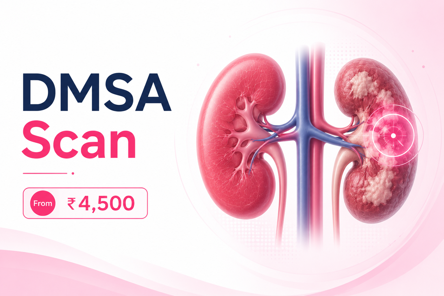
A DMSA scan is the most sensitive test available for detecting kidney scarring. It is not a drainage test and it does not measure GFR. What it does, better than anything else, is show which parts of the kidney cortex are still working and which have been permanently damaged by infection or reflux.
EVE Healthcare offers DMSA scan across Delhi NCR at AERB-licensed nuclear medicine centres from ₹4,500, reported by a qualified nuclear medicine physician. Because the tracer must bind to the renal cortex before imaging, expect to spend three to four hours at the centre. Book on WhatsApp or call +91 9990032078.
What a DMSA Scan Actually Shows
Tc-99m DMSA binds to the tubular cells of the renal cortex and stays there. That is the whole basis of the test. Two to three hours after injection, a gamma camera captures a static image of exactly where functioning cortical tissue exists, and where it does not.
Areas of reduced uptake mean less functioning tissue. Areas of absent uptake mean none at all. From this the physician can identify cortical scarring, acute pyelonephritis, damage from vesicoureteric reflux, and congenital abnormalities such as ectopic kidney, horseshoe kidney, or renal agenesis. The software also calculates differential renal function, comparing uptake between the two kidneys.
Understanding a Normal DMSA Scan Report
This is the question most patients arrive with, and few pages answer it clearly.
A normal DMSA report shows differential renal function between 45 and 55 percent for each kidney, with smooth cortical outlines and no photopenic defects. The two kidneys share the work roughly evenly.
Below that, the standard interpretation runs as follows. Function of 40 to 44 percent is mildly reduced. 10 to 39 percent is substantially impaired. Below 10 percent the kidney is considered non-functioning. Separately, a difference of 10 percent or more between the two kidneys is treated as a significant loss of function, allowing for measurement error of up to 10 percent.
A renal scar is formally defined as a loss of functional tissue in at least two directions, seen as a persistent wedge-shaped defect months after infection. Your treating doctor interprets these numbers alongside your clinical history, because the same figure means different things in a child with reflux and an adult with recurrent pyelonephritis.
Why Ultrasound Cannot Replace It
Ultrasound is the first scan most patients get, and it misses most scars. In a study of 285 renal units, ultrasound identified parenchymal defects in 14 percent of kidneys while DMSA scintigraphy identified them in 31 percent, giving ultrasound a sensitivity of roughly 36 percent against DMSA.
Put plainly, an ultrasound that reports normal kidneys does not rule out scarring. This is why paediatric nephrologists insist on DMSA after febrile urinary tract infection rather than relying on ultrasound alone.
Who Needs a DMSA Scan?
The most common indication is a child with recurrent or febrile urinary tract infection, where the question is whether infection has left permanent cortical damage. Every child in whom vesicoureteric reflux is confirmed carries a meaningful risk of scarring, and DMSA is the only reliable way to establish it.
Beyond that, DMSA is used to confirm acute pyelonephritis during infection, to follow up known scarring over time, to assess ectopic or horseshoe kidneys and suspected renal agenesis, and to establish differential function where a static, structural answer is what the clinician needs rather than a dynamic one.
Scarring matters because it is a recognised cause of hypertension, and extensive scarring can progress to chronic renal impairment. A DMSA result frequently changes management, prompting different antibiotics, treatment of bladder dysfunction, or surgical intervention to prevent further damage.
Timing: When the Scan Should Be Done
Timing is not a detail. It determines whether the result is meaningful.
To detect permanent scarring, the scan is performed four to six months after a febrile urinary tract infection. Done earlier, acute inflammation appears as reduced uptake and is indistinguishable from a scar, producing a false result that can lead to unnecessary intervention.
To confirm acute pyelonephritis during the infection itself, DMSA can be done immediately, where reduced uptake indicates active inflammation. These are two different questions and two different timings. Your doctor is asking one or the other.
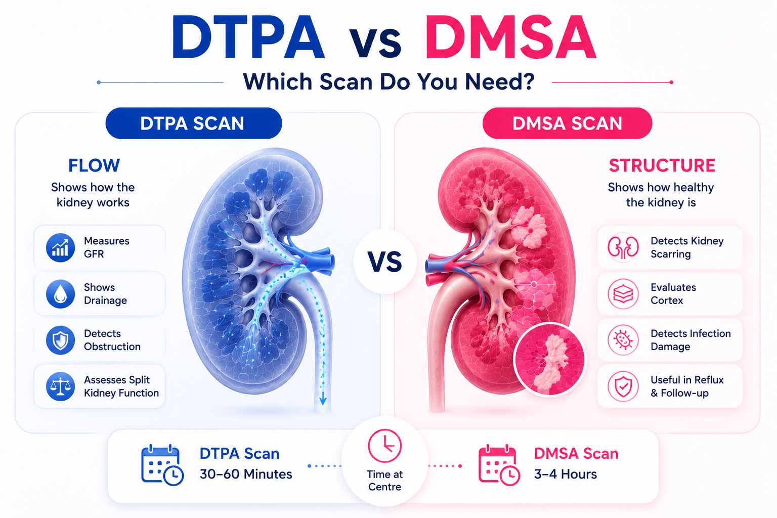
DMSA vs DTPA — Which Scan Do You Need?
| DMSA | DTPA | |
|---|---|---|
| Type | Static | Dynamic |
| Tracer behaviour | Binds to cortical tubules | Cleared by glomerular filtration |
| Measures GFR? | No | Yes, directly |
| Shows | Cortical scarring, pyelonephritis | Blood flow, GFR, drainage, obstruction |
| Duration | 3 to 4 hours (2-3 hr uptake + imaging) | 30 to 60 minutes |
| Ordered for | Recurrent UTI, reflux, scarring | Hydronephrosis, obstruction, transplant, donor workup |
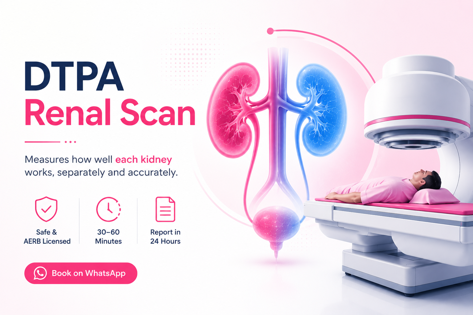
They answer different questions and are not interchangeable. If your prescription says DMSA, a DTPA renal scan will not show cortical scars. Always follow your specialist's prescription.
DMSA Scan Cost in Delhi NCR
| Scan | EVE Healthcare | Typical Delhi range | CGHS (NABH) |
|---|---|---|---|
| DMSA Scan | from ₹4,500 | ₹4,000 – ₹8,000 | ₹3,500 – ₹5,000 |
| DMSA with SPECT | ₹5,500 – ₹9,000 | ₹5,000 – ₹10,000 | on approval |
The spread across Delhi reflects hospital overheads rather than scan quality. What determines whether your report is usable is the AERB licence and whether a qualified nuclear medicine physician interprets it. EVE Healthcare partner centres are AERB-licensed, with CGHS and ECHS cashless available at empanelled centres.
Some centres add SPECT imaging, which gives a volumetric estimate of defect size and is more accurate than planar imaging for assessing how much tissue has been lost. Ask whether it is included if your doctor has requested it.
How to Prepare and What to Expect
There is very little preparation. Eat normally, since no fasting is required, and stay well hydrated. Continue your regular medications. Bring your specialist's prescription, as AERB-licensed centres cannot proceed without one, and tell the physician if you are or may be pregnant, or breastfeeding.
On the day, a small IV line is placed and the Tc-99m DMSA tracer is injected. Then you wait. The tracer needs two to three hours to bind to the renal cortex, and imaging before that produces a poor study. Bring something to occupy the time, and expect to be at the centre for three to four hours in total.
Imaging itself takes thirty to forty-five minutes, with static images taken from several angles. In children, the dose is adjusted by age and weight. Afterwards you resume normal activity immediately, drinking plenty of water so the tracer clears in urine.
Is a DMSA Scan Safe?
Yes, including in children, where it is routinely performed. The Tc-99m dose is small, comparable to a few months of natural background radiation, and is adjusted for a child's age and weight. There is no iodinated contrast dye, so there is no risk of contrast nephropathy. Side effects are rare. It is avoided in pregnancy.
What to Confirm Before Booking
Confirm the centre holds a valid AERB licence, mandatory for every nuclear medicine scan in India, and that a qualified nuclear medicine physician reads and signs the report rather than a technician. Ask whether SPECT is included if your doctor requested it, and confirm tracer availability on your appointment date, since Tc-99m has a half-life of roughly six hours and is prepared in batches rather than kept in stock. Check report turnaround, and if you are a CGHS or ECHS beneficiary, confirm empanelment for this specific scan rather than assuming general empanelment covers it.
Related Scans
Book your DMSA scan — WhatsApp +91 9990032078 or call. AERB-licensed nuclear medicine centres across Delhi NCR · From ₹4,500 · Reports by nuclear medicine physicians.
.png)
-(1).png)
.png)