.png)
PSMA PET CT Scan Cost in Delhi NCR 2026
| Tracer Type | EVE Partner Centre | Private Hospital | CGHS Rate (NABH) |
|---|---|---|---|
| Ga-68 PSMA PET CT | ₹16,000 – ₹25,000 | ₹30,000 – ₹50,000 | ₹12,750 – ₹15,000 |
| F-18 PSMA (PyL/DCFPyL) | ₹22,000 – ₹35,000 | ₹35,000 – ₹55,000 | ₹12,750 – ₹15,000 |
Same-week availability at AERB-licensed nuclear medicine centres across Delhi, Noida and Gurgaon. Book via EVE Healthcare — WhatsApp +91-9990032078
So… What Exactly is a PSMA PET CT Scan?
Let's start simple. If you or someone you love has been diagnosed with prostate cancer, your oncologist may have mentioned a "PSMA PET CT scan." It sounds intimidating, but the idea behind it is actually quite elegant.
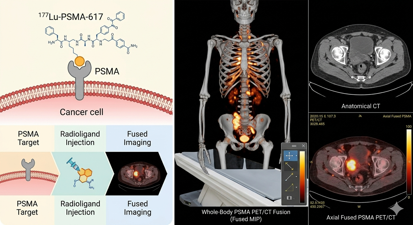
PSMA stands for Prostate-Specific Membrane Antigen — a protein that exists in large amounts on the surface of prostate cancer cells. Think of PSMA as a unique docking station that cancer cells display on their surface. Normal cells have very little of it; prostate cancer cells are practically covered in it.
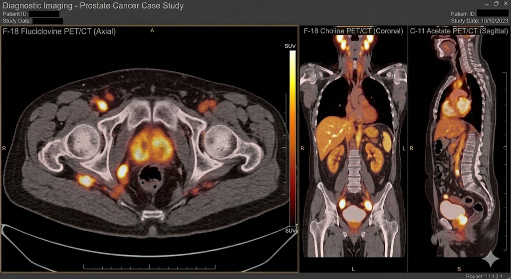
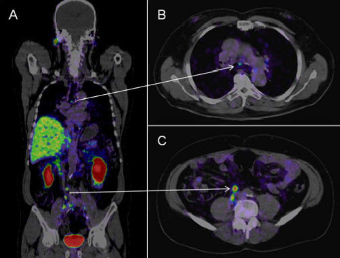
A PSMA PET CT scan uses a mildly radioactive tracer — most commonly Gallium-68 (Ga-68) — that is designed to seek out and lock onto these PSMA proteins. Once the tracer is in your body, the PET (Positron Emission Tomography) scanner detects where it has accumulated, and the CT portion creates a detailed anatomical map. Together, they produce a full-body picture of where prostate cancer cells are hiding.
"PSMA PET CT is like giving your doctors a GPS that tracks cancer cells directly — not just shadows or swellings, but the actual biology of the disease."
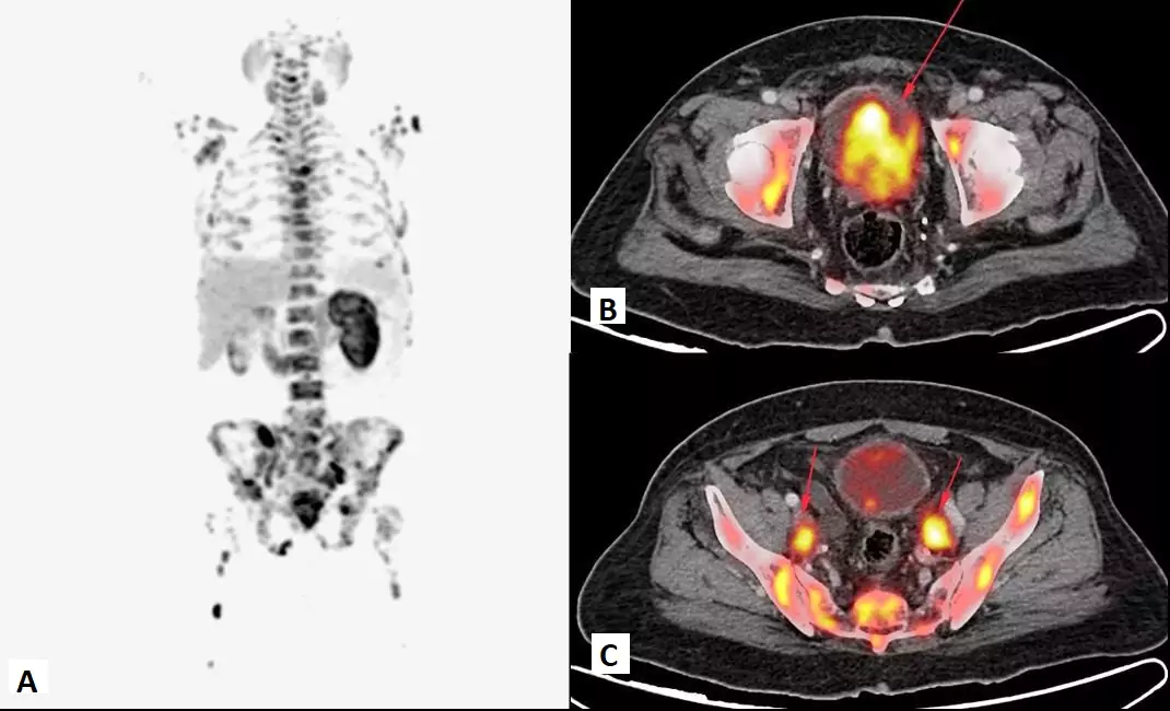
It is the most sensitive tool available today for detecting prostate cancer spread — outperforming older techniques like bone scans and conventional CT by a significant margin.
- 95% Sensitivity for detecting spread
- 40% More accurate than CT + bone scan
- 1 Single scan — whole-body assessment
- ~3 hrs Total procedure time
How Does a PSMA PET CT Actually Work?
Step 1 — Preparing the Radiotracer
Gallium-68 (or sometimes F-18 PSMA) is attached to a molecule specifically engineered to bind to PSMA receptors. This radiotracer is produced fresh on the day of your scan — it has a short half-life, so timing matters.
The radioactivity used is very low — roughly equivalent to what you would receive from a CT scan alone. It leaves your body naturally within hours via urine.
Step 2 — Injection into Your Bloodstream
The tracer is injected through a simple IV line, just like a blood test. It enters your bloodstream and immediately begins travelling throughout your body, seeking out cells that express PSMA proteins.
You will not feel anything different — no sensation, no heat, no discomfort from the tracer itself. Some patients experience mild discomfort at the injection site, which is entirely normal.
Step 3 — The Uptake Wait (45 to 60 minutes)
After injection you will wait 45 to 60 minutes while the tracer binds to any PSMA-expressing cancer cells in your body. During this time you will rest quietly — ideally lying down. Avoid walking around unnecessarily. You will also be asked to drink water and urinate before the scan to clear tracer from your bladder.
Step 4 — The Actual Scan (20 to 30 minutes)
You lie still on a comfortable table that slowly moves through the PET CT scanner — a large, donut-shaped machine. The PET component detects gamma rays emitted by the tracer wherever it has accumulated. The CT simultaneously captures your anatomy in detail.
The scanner is quiet and open — far less claustrophobic than an MRI. Most patients find it quite manageable.
Step 5 — Image Fusion and Reporting
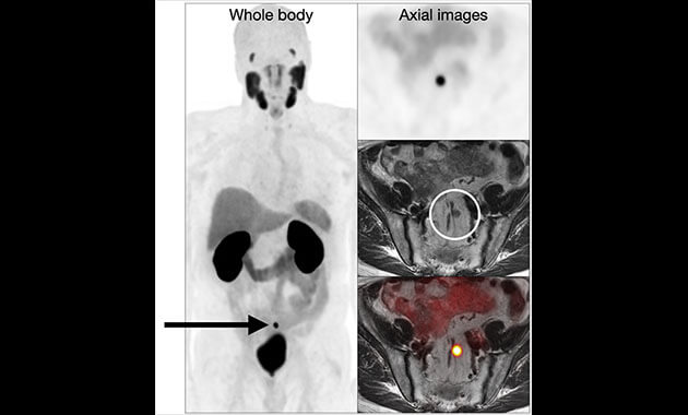
The PET and CT images are fused by sophisticated software and then reviewed by a nuclear medicine physician. Areas of abnormally high PSMA uptake light up on the PET scan, and their precise location is confirmed on the CT.
The radiologist writes a detailed report indicating the location, intensity and nature of any detected lesions. This report goes to your oncologist, who uses it to plan your treatment.
Report ready within 24 hours at EVE partner centres.
Who Should Get a PSMA PET CT Scan?
PSMA PET CT is not a screening test for the general population. It is specifically used in men who already have a prostate cancer diagnosis or strong suspicion.
✅ Staging at initial diagnosis High-risk prostate cancer (high PSA, Gleason score ≥8, or bulky tumour). Needed to see if cancer has spread before deciding on surgery or radiation.
✅ Biochemical recurrence (BCR) PSA rising again after prostatectomy or radiation therapy, but conventional scans show nothing. PSMA PET can detect recurrence at PSA levels as low as 0.2 ng/mL.
✅ Pre-surgical planning Surgeons use PSMA PET to identify lymph node involvement before radical prostatectomy, enabling more targeted node dissection.
✅ Treatment response monitoring Tracking how well hormonal therapy or PSMA-targeted therapy (Lu-177 PSMA) is working.
✅ Before PSMA-targeted therapy (Lutetium-177) A diagnostic PSMA PET scan is mandatory to confirm your tumours express PSMA before this treatment.
❌ Not for general screening Not recommended in men without a prostate cancer diagnosis or with low-risk, localised disease managed conservatively on active surveillance.
PSMA PET CT vs Conventional Scans
| Feature | Bone scan + CT | MRI | PSMA PET CT |
|---|---|---|---|
| Detection basis | Anatomy and bone turnover | Soft tissue anatomy | Cancer cell biology — PSMA protein |
| PSA detection threshold | Above 20 ng/mL realistically | Above 1 ng/mL (local only) | As low as 0.2 ng/mL |
| Whole-body coverage | Two separate scans needed | No — region specific | Yes — head to toe in one |
| Sensitivity | 40 to 60% | 60 to 70% (local) | 90 to 95% |
| Soft tissue lesions | Poor | Good (prostate only) | Excellent (everywhere) |
| Changed treatment plan | 28% of cases | Limited data | 54% of cases |
| False positives | High — fractures, arthritis | Moderate | Very low |
The landmark proPSMA trial (2020, Australia) showed PSMA PET CT was 27% more accurate than conventional imaging for initial staging of high-risk prostate cancer. It detected nearly twice as many pelvic nodal metastases and directly changed treatment plans in 28% more patients.
What to Expect on Scan Day
Morning — Fasting (if required) Unlike FDG PET CT, PSMA PET CT does not require fasting for most centres. Avoid intense exercise 24 hours before — muscle uptake can interfere with image quality. Continue your regular medications unless told otherwise.
Arrival — Registration and prep (15 minutes) You will check in, have your blood sugar checked, and confirm your medical history. A simple IV line is placed — similar to a blood draw.
Tracer injection and rest period (60 minutes) The Ga-68 PSMA tracer is injected. You will rest quietly in a comfortable room. Drink the water provided — it helps flush tracer from your kidneys and bladder.
The scan itself (30 minutes) You lie on the scanner table, arms at your sides or above your head. The machine is quiet and open. Breathe normally. Stay as still as possible. The table moves slowly through the scanner ring.
Done — Back to normal No recovery time needed. You can drive, eat and go about your day. Drink extra water for 24 hours to flush the tracer. Avoid prolonged close contact with pregnant women and young children for about 6 hours as a simple precaution.
Results — 24 hours later at EVE partner centres The nuclear medicine physician analyses the images and writes the report. Your oncologist will receive it and discuss the findings with you.
Understanding Your PSMA PET CT Report
PSMA-RADS Scoring System (1 to 5)
Many centres use this standardised scoring system to report findings:
- 1 — No lesion detected: Highly unlikely to be prostate cancer
- 2 — Probably not cancer: Low suspicion finding — typically benign
- 3 — Equivocal: Neither typical nor atypical — may need further investigation
- 4 — Probably prostate cancer: High confidence of cancer involvement
- 5 — Almost certainly cancer: Very intense PSMA uptake, classic appearance
Other Terms in Your Report
SUVmax — Maximum Standardized Uptake Value. A measure of how intensely the tracer accumulated. Higher = more PSMA expression = more suspicious.
Avid lesion — A spot that actively took up the tracer. "PSMA-avid" means it is lighting up on the scan.
Oligometastatic disease — A small number of metastatic lesions (typically 1 to 5). Important because it may be treatable with targeted radiation (SABR/SBRT).
Physiological uptake — Normal tracer accumulation in organs like kidneys, salivary glands and lacrimal glands. Not concerning.
Insurance and CGHS Coverage
PSMA PET CT is increasingly covered by private health insurance and CGHS when appropriately indicated — biochemical recurrence, high-risk staging. Always obtain a proper referral letter from your oncologist specifying the clinical indication — it significantly improves your approval chances.
CGHS rate: ₹12,750 to ₹15,000 at NABH-accredited empanelled centres.
Questions to Ask Your Oncologist Before Booking
- Am I a good candidate for PSMA PET CT, and why specifically do you recommend it for my case?
- Which tracer will be used — Ga-68 PSMA or F-18 PSMA (DCFPyL/PyL)?
- Should I continue or temporarily stop hormonal therapy before the scan?
- How will the results change my treatment plan? What are the possible next steps?
- Will insurance or CGHS cover this, and can you provide a referral letter?
Book PSMA PET CT Scan in Delhi NCR
EVE Healthcare partner AERB-licensed nuclear medicine centres offer Ga-68 PSMA PET CT from ₹16,000 and F-18 PSMA from ₹22,000 across Delhi NCR with same-week availability and reports within 24 hours.
Book online at eve-healthcare.com or WhatsApp +91-9990032078 to confirm tracer availability and book your appointment.
.png)
-(1).png)