
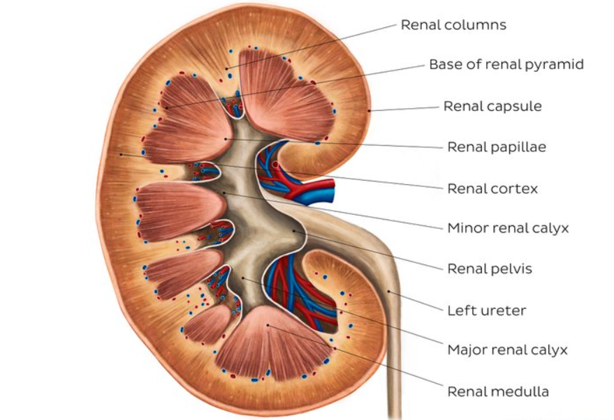
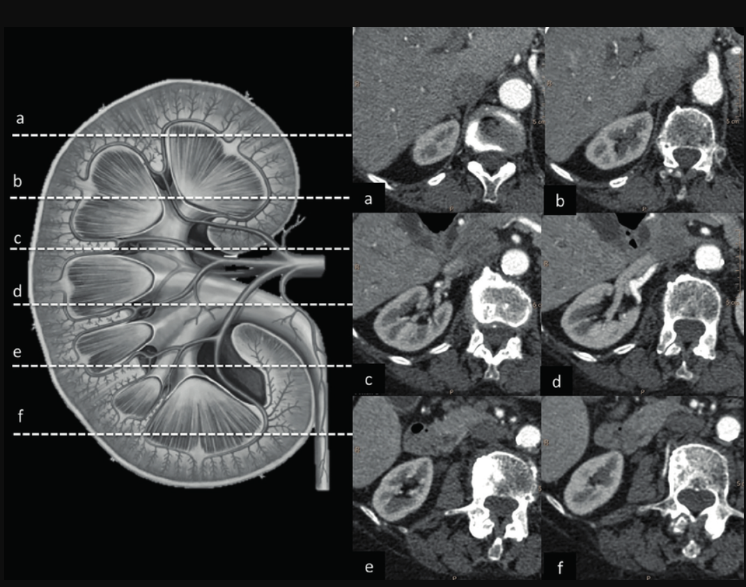
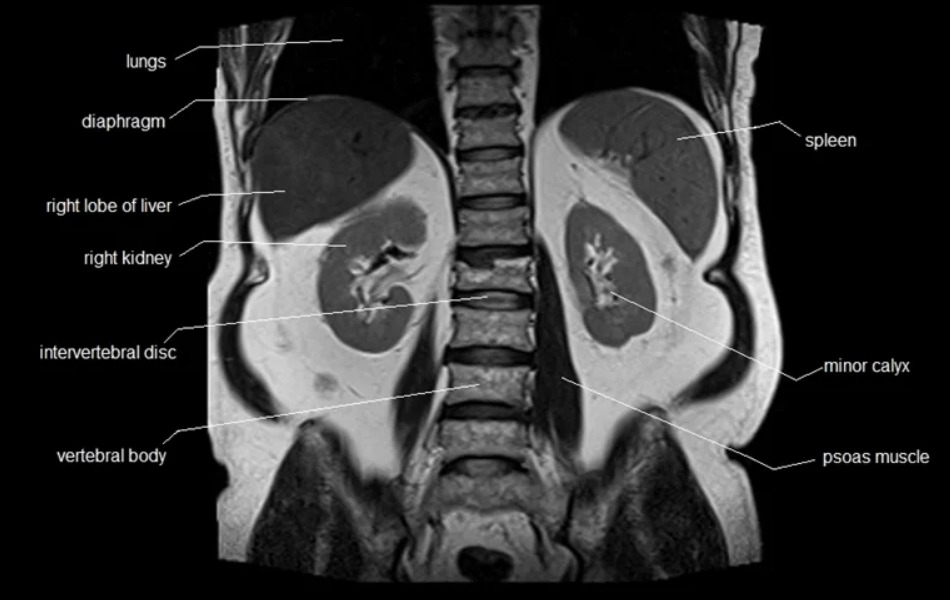
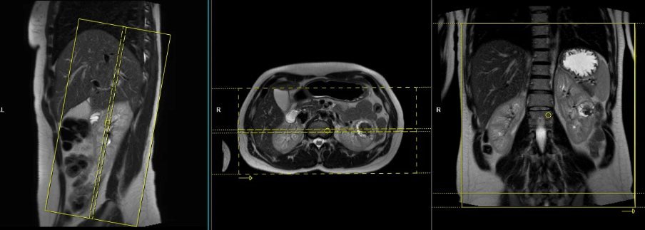
If your doctor has recommended an MRI of the kidneys — or your CT scan has found a renal mass that needs further characterisation — this guide explains exactly what MRI shows that CT and ultrasound cannot, what it costs in Delhi NCR in 2026, and how to book.
Book MRI kidney scan at NABL-accredited diagnostic centres across Delhi NCR — WhatsApp +91-9990032078
MRI Kidney Scan Cost in Delhi 2026
| Scan Type | Standalone Lab | Private Hospital | CGHS Rate (NABH) |
|---|---|---|---|
| MRI Abdomen plain (includes kidneys) | ₹4,500 – ₹9,000 | ₹10,000 – ₹22,000 | ₹4,500 |
| MRI Abdomen with contrast (CEMRI) | ₹6,000 – ₹12,000 | ₹14,000 – ₹28,000 | ₹6,000 |
| MRI Urography (MRU) | ₹5,000 – ₹10,000 | ₹12,000 – ₹25,000 | ₹5,000 |
| MRI Renal Angiography (MRA) | ₹6,000 – ₹12,000 | ₹14,000 – ₹28,000 | ₹6,000 |
Book via EVE Healthcare — WhatsApp +91-9990032078 for same-day appointments across South Delhi, Noida, Gurgaon and Ghaziabad.
What is an MRI of the Kidneys?
Magnetic Resonance Imaging (MRI) of the kidneys uses powerful magnetic fields and radio waves to produce detailed images of kidney structure, masses, cysts and vascular supply — without ionising radiation. This makes MRI particularly valuable for patients who need repeated imaging, children, pregnant women and patients with contrast allergy who cannot safely receive CT contrast dye.
Unlike a CT scan which uses X-ray radiation, or an ultrasound which has limited resolution for small lesions and cannot assess function, MRI provides multiparametric imaging — multiple sequences that together characterise tissue composition, vascularity and function in a single examination.
When is MRI of the Kidneys Ordered?
Renal Mass Characterisation — Most Common Indication
When a kidney mass or lesion is found on ultrasound or CT but its nature is uncertain, MRI is the investigation of choice. CT can detect masses but often cannot distinguish between benign and malignant lesions with confidence. MRI's multiparametric sequences — T1, T2, diffusion-weighted imaging (DWI) and dynamic contrast enhancement — provide tissue characterisation that CT cannot.
What MRI can determine about a renal mass:
- Whether it is a simple cyst (benign, no treatment needed) or a complex cyst requiring follow-up or surgery
- Whether a solid mass enhances with contrast — enhancement is the key feature distinguishing renal cell carcinoma from benign angiomyolipoma or oncocytoma
- The Bosniak classification of cystic lesions (I to IV) — which determines management
- Whether fat is present within a mass — diagnosing angiomyolipoma without biopsy
Renal Cell Carcinoma — Staging
MRI accurately stages renal cell carcinoma (RCC) — the most common kidney cancer:
- Tumour size and exact location within the kidney
- Whether the tumour has invaded the renal vein or inferior vena cava — critical for surgical planning
- Whether perinephric fat or adjacent organs are involved
- Regional lymph node status
Polycystic Kidney Disease (PKD) — Monitoring
MRI is preferred over CT for long-term monitoring of autosomal dominant polycystic kidney disease (ADPKD) because it avoids repeated radiation exposure over years of follow-up. It measures total kidney volume — the primary endpoint for disease progression and treatment response monitoring.
Renal Artery Stenosis — MR Angiography
MR angiography (MRA) of the renal arteries assesses narrowing or occlusion of the arteries supplying the kidneys — a cause of renovascular hypertension and renal dysfunction. It is a non-invasive alternative to conventional catheter angiography.
Congenital Kidney Abnormalities
MRI excels at mapping complex congenital anomalies — horseshoe kidney, duplex collecting system, ectopic kidney — where the spatial relationships between structures are important for surgical planning.
Renal Infections and Abscesses
When ultrasound is inconclusive for suspected pyelonephritis or renal abscess, MRI provides detailed assessment of the extent of infection and any associated complications.
MR Urography — Urinary Tract Assessment
MR urography (MRU) provides detailed imaging of the entire urinary collecting system — kidneys, ureters and bladder — without radiation. It is particularly useful for evaluating hydronephrosis, ureteral obstruction and congenital urinary tract anomalies in children.
Kidney Function Assessment
Functional MRI sequences assess perfusion and filtration at a tissue level — providing information about individual kidney function that cannot be obtained from standard blood tests alone.
MRI vs CT vs Ultrasound for Kidneys
| MRI | CT Scan | Ultrasound | |
|---|---|---|---|
| Radiation | None | Yes | None |
| Renal mass characterisation | Gold standard | Good — first line | Limited |
| Small lesion detection | Excellent | Good | Limited |
| Renal vein invasion | Excellent | Good | Poor |
| Bosniak classification | Excellent | Good | Not applicable |
| Kidney stones | Poor | Gold standard | Moderate |
| Renal artery assessment | Excellent (MRA) | Good (CTA) | Limited |
| Function assessment | Yes | No | No |
| Best for | Complex masses, staging, follow-up | Initial assessment, stones | First-line screening |
Key point: Ultrasound is the first-line investigation. CT is ordered when ultrasound finds something abnormal. MRI is ordered when CT findings need further characterisation — particularly for renal masses.
What Does MRI of the Kidneys Show?
Tumours and Masses
MRI provides multiparametric tissue characterisation — T2 signal, enhancement pattern on dynamic contrast series, and diffusion restriction — that together allow highly accurate differentiation between malignant and benign renal masses. It assesses exact tumour size, location, vascular invasion and surgical margins.
Bosniak Classification of Cystic Lesions
The Bosniak classification system grades renal cystic lesions from I to IV based on their imaging features:
| Bosniak Class | Features | Malignancy Risk | Management |
|---|---|---|---|
| I | Simple cyst — thin wall, no septa | 0% | No follow-up needed |
| II | Minimal complexity — thin septa, small calcification | 0 – 5% | No follow-up needed |
| IIF | Increased complexity — multiple thin septa | 5 – 15% | Follow-up imaging |
| III | Thickened irregular septa or wall | 50 – 55% | Surgery or biopsy |
| IV | Solid enhancing components | 85 – 100% | Surgery |
Hydronephrosis
MRI detects the buildup of urine in the kidneys due to blockages in the ureters or bladder from stones, tumours or congenital abnormalities. MR urography maps the complete collecting system.
Vascular Abnormalities
Renal artery stenosis, renal vein thrombosis and other vascular abnormalities are clearly demonstrated on MRA sequences.
Infections and Abscesses
MRI accurately delineates the extent of renal and perirenal infection and distinguishes organised abscess from inflammatory phlegmon — helping guide drainage decisions.
How to Prepare for MRI of the Kidneys
Plain MRI kidney (without contrast):
- Fast for 4 to 6 hours — fasting reduces bowel motion artefacts that can degrade abdominal image quality
- Water is allowed
- Remove all metal objects and jewellery
MRI kidney with gadolinium contrast:
- Fast for 4 to 6 hours
- Bring a recent creatinine blood test result — gadolinium contrast requires adequate kidney function (eGFR above 30)
- Inform the technician of any kidney disease, previous gadolinium reaction, or dialysis
- Remove all metal objects
For all MRI:
- Inform the team of any metal implants before booking
- The scan takes 30 to 60 minutes depending on sequences required
- Breath-hold instructions will be given during the scan — practice taking a deep breath and holding for 15 to 20 seconds
What to Expect During the Procedure
One — You lie on your back on the MRI table with a surface coil placed over your abdomen
Two — The table slides into the MRI scanner — open at both ends, not a closed tunnel
Three — The machine makes loud knocking sounds — earplugs or headphones are provided
Four — You will be asked to hold your breath for 15 to 20 seconds at multiple intervals during the scan — this eliminates breathing motion from the images
Five — If contrast is required — a small IV line in your arm delivers gadolinium during the scan
Six — After the scan — resume normal activities immediately. Drink plenty of water if contrast was used.
Total appointment time: 60 to 90 minutes including registration and preparation.
Risks and Limitations
Gadolinium contrast: Generally very safe. Rare allergic reactions occur. In patients with severely reduced kidney function (eGFR below 30), gadolinium carries a very small risk of nephrogenic systemic fibrosis — inform the radiologist of any kidney disease before contrast is administered.
Claustrophobia: The MRI scanner is open at both ends. Most patients tolerate it well. Wide-bore 1.5T machines (70cm opening) are available at several EVE partner centres for patients who are concerned about confined spaces.
Metal implants: Some implants are not MRI-compatible. Always inform the booking team of any implants — pacemakers, cochlear implants, certain aneurysm clips — before scheduling.
Kidney stones: MRI is not the recommended investigation for kidney stones — CT KUB is the gold standard. If kidney stones are the primary concern, CT KUB is the correct test.
Book MRI Kidney Scan in Delhi NCR
EVE Healthcare partner NABL-accredited diagnostic centres offer MRI abdomen including dedicated renal imaging across Delhi NCR starting from ₹4,500 with same-day availability and reports within 4 to 6 hours.
Book online at eve-healthcare.com or WhatsApp +91-9990032078 to confirm same-day appointments across South Delhi, Noida, Gurgaon and Ghaziabad.
.png)
.png)
.png)
-(1).png)
-(1)-(1).png)
-(1).png)