
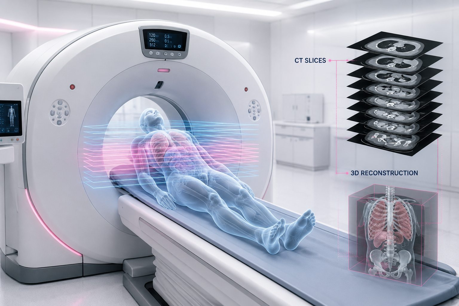
When a CT scanner is described as "64-slice" or "256-slice," that number is one of the most misunderstood specifications in medical imaging. It is marketed as a measure of quality. It is really a measure of coverage and speed. This guide explains what a slice actually is, what the number means, and when it matters to you as a patient, without the sales pitch.
What Is a Slice?
A slice is a single cross-sectional image of the body, a thin layer viewed as if the body had been cut across and you were looking at the cut face. A CT scanner rotates an X-ray tube around you while detectors on the opposite side record how much radiation passes through. A computer reconstructs those measurements into one cross-sectional image: one slice.
The scanner captures many of these along the length of the body, and stacking them builds a full three-dimensional picture. Scroll through a CT study and you are moving through the stack, one slice at a time.
What Does the Slice Number Actually Mean?
The number, 16, 64, 128, 256, refers to the rows of detectors arranged along the length of the patient, and therefore how many slices the scanner captures in a single rotation of the gantry.
A 64-slice scanner has 64 detector rows recording simultaneously. Each gantry rotation takes roughly 0.27 to 0.5 seconds, and because the table moves continuously through the ring, the scan traces a smooth helix rather than separate rings. This is helical, or spiral, CT.
More rows means more of the body captured per rotation. That is the entire advantage: coverage and speed, not inherent image quality.
How Slice Count Affects Scan Time
The practical impact is dramatic. A 64-slice scanner covers four times the anatomy per rotation of a 16-slice, so it needs a quarter of the rotations for the same region.
Concretely: a 64-slice scanner can image 500 mm of anatomy at 1 mm slices in about 7.8 seconds. A 16-slice scanner covering the same region takes about 31 seconds.
That speed is why higher slice counts matter for specific jobs: freezing a beating heart in CT coronary angiography, scanning a trauma patient in seconds, or imaging someone who cannot hold their breath. For a routine head or abdomen scan on a cooperative patient, the extra speed changes nothing about the diagnosis.
The Evolution of Slice Counts
Multislice CT began in 1998 with 4-slice scanners. 16-slice arrived in 2002, and 64-slice from 2005 became the workhorse of modern radiology. Today the commonly available counts are 16, 32, 40, 64 and 128, with high-end systems offering 256, 320 and even 640 slices. Four and eight-slice machines have largely been withdrawn.
A crucial and rarely stated point from the imaging literature: beyond 16-slice, z-axis resolution and scan quality are essentially identical. For most examinations, the gain from higher slice counts is scan time, which was already extraordinarily short at 16-slice. The exceptions are CT angiography and cardiac work, which genuinely benefit from the added speed and coverage.
Slice Thickness — The Specification That Actually Affects Your Images
Here is the distinction most people miss. Slice count is how many slices per rotation. Slice thickness is how thick each slice is. Thickness, not count, is what determines the fine detail in your images.
Slice thickness on modern scanners ranges from about 0.625 mm to 5 mm. It behaves like pixel size in a photograph: the thinner the slice, the higher the spatial resolution and the finer the detail.
But there is a fundamental trade-off:
Thinner slices = better spatial resolution + more radiation dose + larger files + more images for the radiologist to review.
Thicker slices = more coverage per rotation + less image noise + small structures blurred.
Thin vs Thick Slices — When Each Is Used
Thin slices, around 0.625 to 1 mm, are chosen where fine detail is the whole point: HRCT of the lungs, temporal bone imaging, CT angiography, and hunting for small lesions or subtle fractures.
Thick slices, around 3 to 5 mm, are chosen for routine survey imaging, where they reduce noise, cut the number of images, and are perfectly adequate to answer the clinical question.
Radiologists routinely acquire thin slices and then reconstruct thicker ones from the same data, getting both detail and a manageable review set from a single scan. The choice is clinical, not a fixed property of the machine.
How the Detector Actually Works
A subtle but important detail: many scanners have more physical detector rows than their slice count. A 16-slice system, for instance, has more than 16 rows. The narrow rows in the centre acquire thin slices; the wider outer rows acquire thick ones.
When thin slices are selected, only the narrow central detectors are used, so a shorter length of the patient is imaged per rotation. When thicker slices are acceptable, the full detector width is used and more anatomy is covered at once. This is the hidden reason a high-detail thin-slice scan of a small area can take a similar time to a coarse scan of a large one.
From Raw Data to the Image You See
Each rotation produces thousands of attenuation measurements, raw data called a sinogram. A reconstruction algorithm, either filtered back-projection or the more modern iterative reconstruction, converts this into the pixel image on screen. A standard CT slice is a 512 by 512 grid of pixels.
Each pixel's brightness is a Hounsfield Unit (HU), a calibrated density value: air is around -1000 HU, water is 0 HU, soft tissue +20 to +80 HU, and bone +400 to +1000 HU. This is why a radiologist can distinguish a fluid-filled cyst from a solid mass by its density alone, and why they adjust the "window" setting to make specific tissues visible.
Does a Higher Slice Count Mean More Radiation?
Counterintuitively, no, not inherently. Slice count does not dictate dose; the protocol does. Higher slice counts, combined with modern reconstruction, can actually lower dose by scanning faster and enabling advanced dose-reduction techniques. What genuinely raises dose is thinner slices, more phases, and unoptimised protocols. A well-run scan on a 256-slice machine can deliver less radiation than a poorly optimised scan on an older one.
So Which Slice Count Do You Actually Need?
For the vast majority of scans, 64-slice is more than enough, and 16-slice is fully diagnostic for routine head, chest and abdomen imaging. A higher count genuinely helps for:
- CT coronary angiography and whole-heart imaging
- Trauma, where speed is critical
- Patients who cannot hold their breath or stay still
- Very young children
For everything else, the single biggest determinant of a useful result is not the scanner's slice count. It is whether the centre is properly licensed and whether a qualified radiologist interprets your scan. A 256-slice scan read poorly is worse than a 64-slice scan read well.
If you are comparing scans, see our guides on CT scan cost in Delhi and, for cardiac imaging where slice count matters most, CT coronary angiography.
Related Guides
- CT Scan Cost in Delhi
- CT Coronary Angiography Cost in Delhi
- CT Scan KUB Cost in Delhi
- MRI vs CT Scan
EVE Healthcare offers CT scans on modern multislice scanners at AERB-licensed centres across Delhi NCR, with reports read by qualified radiologists. WhatsApp +91 9990032078 or call.
-(1).png)
-(1).png)
-(1)-(1).png)
-(1)-(1).png)