
Radiology Deep Dive
If your doctor has prescribed a CT scan and the prescription says "64-slice" or "256-slice" — this guide explains exactly what that means, why it matters for your specific scan, and whether you need to request a particular machine.
How a rotating X-ray beam turns your body into hundreds of paper-thin cross-section images — and why the number of slices changes everything.
640+ Max slices today | 0.5mm Thinnest slice | <5s Full chest scan
The Basics
What is a CT scan "slice"?
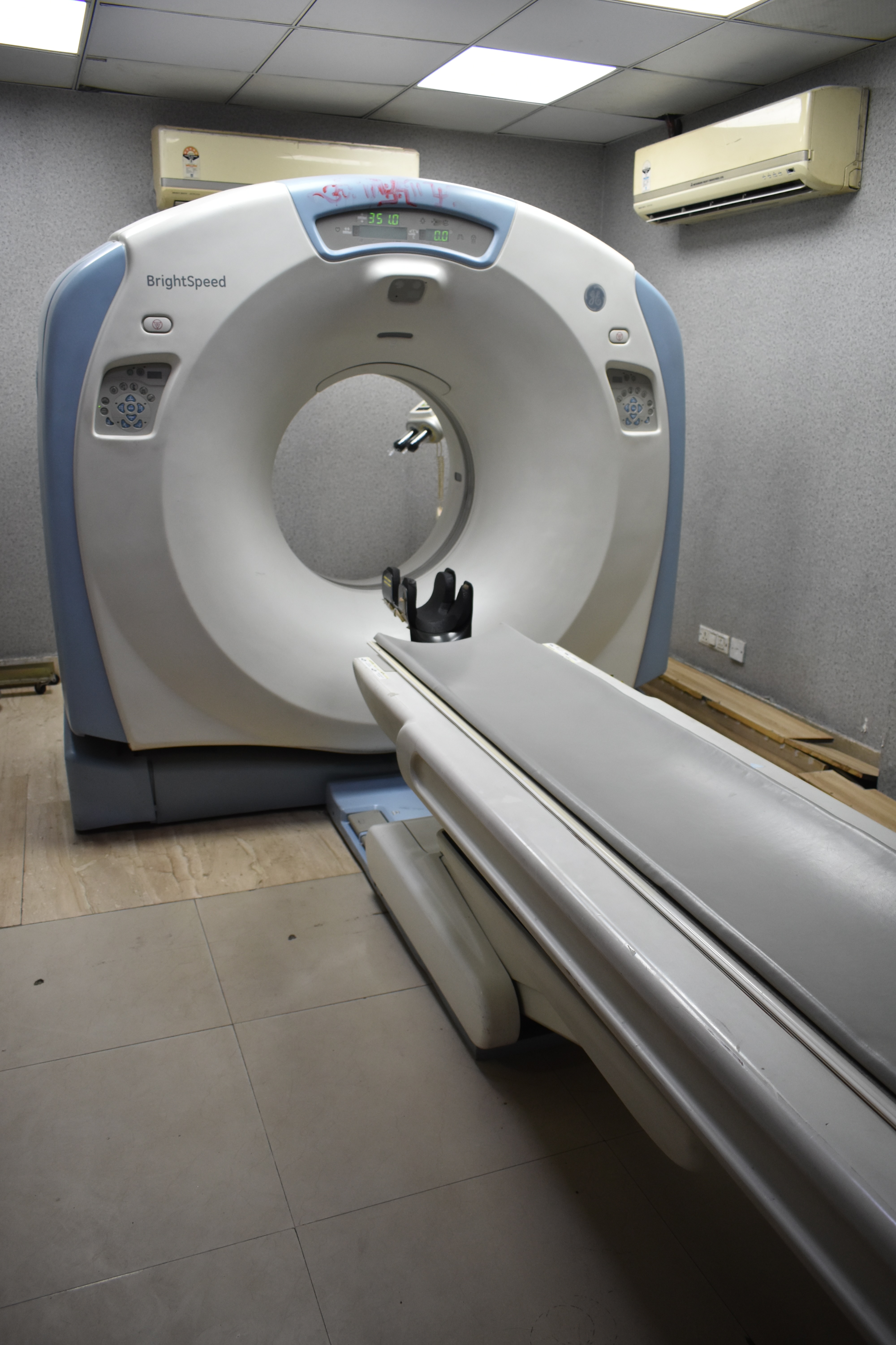
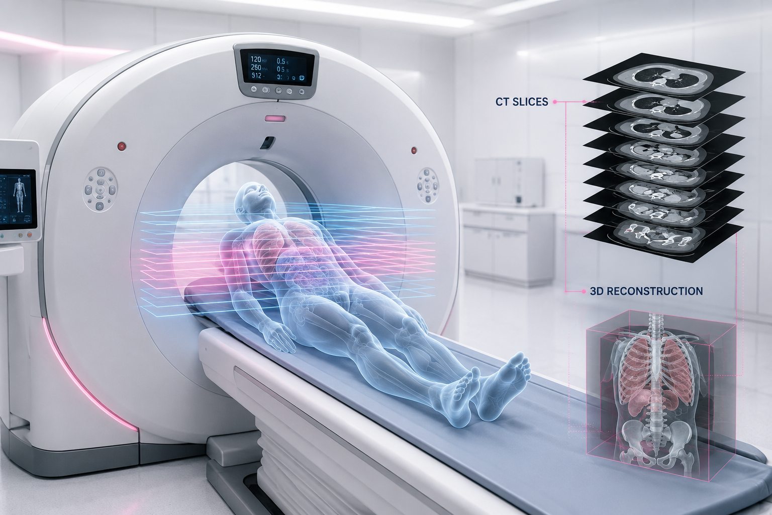
A CT (Computed Tomography) scan works by rotating an X-ray source 360° around your body and capturing thousands of individual X-ray readings. These readings are reconstructed by a computer into flat, 2D cross-sectional images — each image is called a slice.
Think of it like slicing a loaf of bread. Each slice reveals what's inside at that exact depth. Stack all the slices together and you get a full 3D picture of the organ or body part being scanned.
Real Example
A CT scan of the abdomen produces ~150–200 axial slices, each showing a cross-section of your organs at a different height — like viewing your liver, kidneys, and spine level by level from top to bottom.
CT Scan Machine Slices Thickness Explained
Thin vs thick slices — what's the difference?
Slice thickness is the most important parameter in CT imaging. It controls how much anatomy is captured in each image and how fine the detail is.
| Slice Thickness | Use Case |
|---|---|
| 10mm | Very thick — routine chest/abdomen screening |
| 5mm | Standard clinical — most diagnostic CTs |
| 1–2mm | High-resolution — lung, spine detail |
| 0.5–0.625mm | Ultra-thin — cardiac, temporal bone |
Example — Pulmonary Embolism
To detect a small blood clot in a pulmonary artery, radiologists request 1mm slices (CTPA protocol). A 5mm slice would average out the density and miss a small clot entirely. Thinner = more detail, but more radiation dose.
Machine Generations
What does "64-slice" or "128-slice" mean?
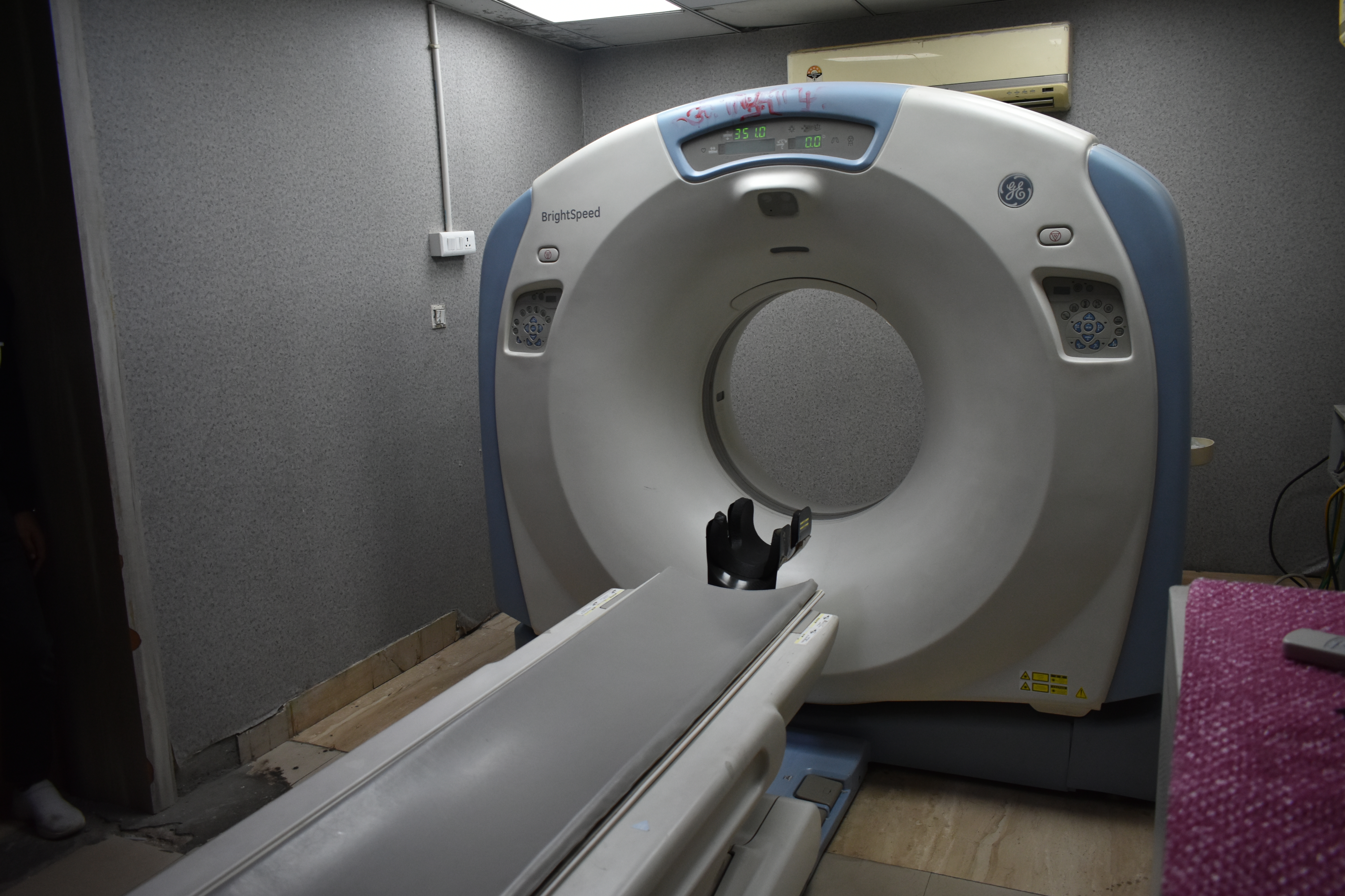
The "slice count" in a CT machine's name refers to the number of detector rows — how many parallel slices it can capture in a single gantry rotation.
| CT Type | Detector Rows | Slices/Rotation | Best For | Speed |
|---|---|---|---|---|
| 4-slice | 4 | 4 | Basic body scans | Slow |
| 16-slice | 16 | 16 | Routine diagnosis | Moderate |
| 64-slice | 64 | 64 | Cardiac, vascular | Fast |
| 128-slice | 64×2 (dual src) | 128 | Cardiac CTA | Very fast |
| 256-slice | 128 | 256 | Whole-heart in 1 beat | Ultra fast |
| 640-slice | 320 | 640 | Dynamic organ imaging | Fastest |
Example — Cardiac CT Angiography
The heart beats 60–100 times/min. A 64-slice CT can freeze the heart in 0.35 seconds per rotation, imaging coronary arteries clearly. A 4-slice CT would produce motion blur — the arteries come out smeared.
How Much Does a Higher Slice CT Scan Cost in Delhi?
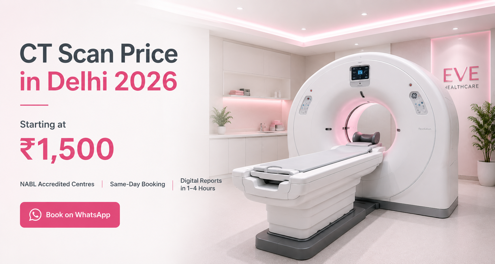
Slice count affects price — but less than most patients expect. For routine scans, a 16-slice and 64-slice CT cost the same at most centres. The premium for higher slice counts applies primarily to cardiac imaging.
| Scanner Type | Common Scans | Price in Delhi |
|---|---|---|
| 16-slice CT | Brain, chest, abdomen | ₹1,500 to ₹3,000 |
| 64-slice CT | Routine and cardiac | ₹2,000 to ₹6,000 |
| 128-slice CT | Complex cardiac and vascular | ₹4,000 to ₹8,000 |
| 256-slice CT | CT Coronary Angiography | From ₹8,000 at EVE partner centres |
For CT Coronary Angiography on a 256-slice scanner, book via EVE Healthcare partner centres across Delhi NCR — WhatsApp +91-9990032078 for same-day confirmation. See our full CT Coronary Angiography Cost in Delhi → guide.
For general CT scan pricing by body part, see our CT Scan Cost in Delhi → guide.
Scan Types
The 3 planes of CT slices
Axial (transverse) Horizontal cross-sections — like slicing a sausage. The default CT plane. Shows left vs right anatomy simultaneously.
Coronal Front-to-back sections dividing front and back half. Great for kidney, colon, and spinal cord views.
Sagittal Left-right sections dividing the body into left and right halves. Essential for spine, brain midline, and pelvis.
MPR (multiplanar) Software reconstructs all 3 planes from raw axial data. Modern CTs acquire axially and reconstruct everything else.
Example — Spinal Disc Herniation
An axial CT slice at the L4-L5 level shows the exact size and direction of disc material pressing on a nerve root. A sagittal reconstruction reveals how many vertebral levels are involved. Both views come from the same scan data.
Clinical Protocols
Real-world slice protocols by body part
| Body Part | Slice Thickness | Why? |
|---|---|---|
| Brain (routine) | 5mm axial | Haemorrhage, infarct detection |
| Brain (pituitary) | 1–2mm coronal | Tiny gland, needs fine detail |
| Chest (lung HRCT) | 1mm axial | Detect early fibrosis, nodules <3mm |
| Abdomen/Pelvis | 3–5mm axial | Organs are large, 5mm is sufficient |
| Coronary CTA | 0.625mm | Coronary arteries are 2–4mm wide |
| Temporal bone (ear) | 0.5–0.6mm | Ossicles are 1–2mm — ultra-thin needed |
| Spine (trauma) | 1.25mm sagittal recon | Fracture lines can be sub-mm |
How It Works
Step-by-step: how a slice is created
- The X-ray tube and detector array rotate 360° around the patient. For a 64-slice CT, 64 rows of detectors record simultaneously.
- Each rotation takes 0.27–0.5 seconds. The table moves continuously (helical/spiral CT), so the scan path is a continuous helix rather than discrete rings.
- Raw data (called "sinograms") is sent to a computer. Thousands of attenuation measurements per slice are collected.
- A reconstruction algorithm (filtered back-projection or iterative) converts sinogram data into a 2D pixel map — the actual image you see.
- Each pixel's brightness = Hounsfield Unit (HU) — air = -1000 HU, water = 0 HU, bone = +400–1000 HU, soft tissue = +20 to +80 HU.
Example — Hounsfield Units in Practice
A liver abscess reads ~0–30 HU (fluid-like), while normal liver is ~55–65 HU, and a fresh bleed is ~50–80 HU. The radiologist adjusts the "window" setting to display these differences clearly.
Key Trade-offs
More slices = better? Not always.
The fundamental trade-off: thinner slices = better spatial resolution + more radiation dose + larger file size + more radiologist workload. A whole-body 0.5mm CT generates 2,000–4,000 images. Clinicians always pick the minimum slice thickness needed for the clinical question.
Thinner slices ✓ Better lesion detection, 3D reconstructions, vascular detail, surgical planning.
Thinner slices ✗ More radiation, more noise per slice, longer processing, larger DICOM files.
Thicker slices ✓ Less noise, lower dose, fewer images, faster reading for routine cases.
Thicker slices ✗ Partial-volume artefact, missed small lesions, poor vascular detail.
Example — Partial Volume Effect
A 5mm slice through a 3mm kidney stone averages the stone's +400 HU with surrounding urine at +5 HU. The final pixel reads ~120 HU — the stone appears softer and may be missed. A 1mm slice captures the stone at its true density.
Latest Technology
Where CT slice technology is today
Modern CT scanners (2020s generation) use photon-counting detectors (PCD-CT), which count individual X-ray photons rather than measuring total energy. This enables sub-0.25mm slices with lower dose and eliminates electronic noise entirely.
The Siemens NAEOTOM Alpha — the first clinical PCD-CT — acquires 120 simultaneous slices at 0.2mm resolution. A chest CT takes under 2 seconds. AI-based iterative reconstruction further reduces noise, allowing dose reductions of 40–80% compared to 2010-era CTs.
Example — Photon Counting CT
With PCD-CT, calcified and non-calcified coronary plaques can be differentiated by their spectral signatures in a single scan — something that previously required two separate scans with different energy settings.
CT slice technology has evolved from 4 slices (1998) to 640+ slices (2020s) — a 160× jump in 25 years. The physics of X-ray attenuation stays the same; what changes is how finely and how fast we sample it.
CT Scan Slice Availability at EVE Healthcare Partner Centres
All EVE Healthcare partner diagnostic centres across Delhi NCR use minimum 64-slice CT scanners for routine scans. 256-slice CT Coronary Angiography is available starting from ₹8,000.
Book online at eve-healthcare.com or WhatsApp +91-9990032078 to confirm which scanner is available at your nearest centre and get same-day appointment confirmation across South Delhi, Noida, Gurgaon and Ghaziabad.
-(1).png)
-(1).png)
-(1)-(1).png)