If your doctor has referred you for a PET CT scan for liver cancer — this page explains the most important clinical distinction in liver cancer imaging: whether you have primary liver cancer (HCC) or liver metastases from another cancer changes which PET CT investigation is appropriate, and what it can reliably show.
FDG PET CT for liver cancer costs ₹9,000–₹14,000 at AERB-licensed nuclear medicine centres in Delhi. For neuroendocrine liver metastases, DOTANOC PET CT costs ₹16,000–₹22,000.
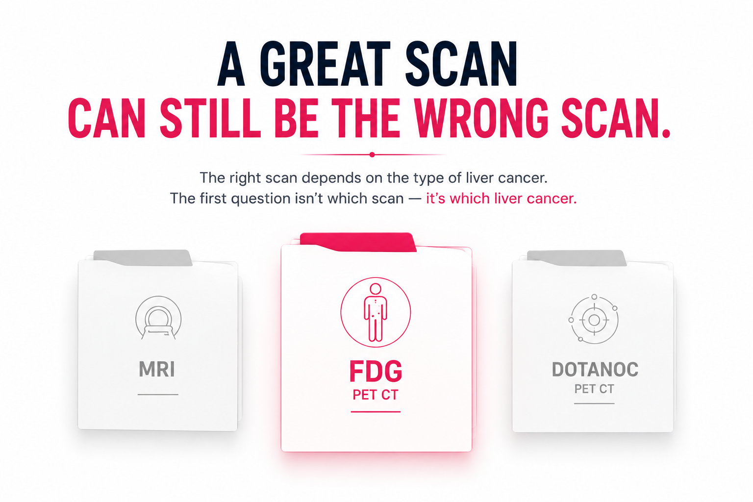
The Single Most Important Distinction — Primary Liver Cancer vs Liver Metastases
This distinction determines which PET CT you need, what it will show, and whether PET CT is even the right investigation for your specific situation.
Primary liver cancer (Hepatocellular Carcinoma — HCC): Cancer that originated in the liver cells. Associated with hepatitis B, hepatitis C, cirrhosis, and alcohol-related liver disease. Very common in India due to high hepatitis B prevalence.
Liver metastases: Cancer from another organ that has spread to the liver — colorectal cancer, breast cancer, lung cancer, neuroendocrine tumours, and others. The primary cancer is elsewhere; the liver is a secondary site.
Why this matters for PET CT:
|
Scenario |
PET CT Performance |
Better Alternative |
|
Primary HCC — well differentiated |
50% false-negative rate |
Contrast-enhanced MRI is superior |
|
Primary HCC — poorly differentiated |
Good FDG avidity |
FDG PET CT useful for staging |
|
Primary HCC — extrahepatic metastases |
85.7% sensitivity — excellent |
FDG PET CT is valuable here |
|
Liver metastases from colorectal cancer |
Good sensitivity |
FDG PET CT + CT colonoscopy |
|
Liver metastases from lung/breast cancer |
Good sensitivity |
FDG PET CT for whole body staging |
|
Neuroendocrine liver metastases (NETs) |
FDG often negative |
DOTANOC/DOTATATE PET CT required |
Primary Liver Cancer (HCC) — The Honest PET CT Picture

HCC is the most common primary liver cancer. India has a high HCC burden due to widespread hepatitis B and C and alcohol-related cirrhosis.
The key clinical limitation of FDG PET CT for HCC:
The false-negative rate of FDG PET CT is almost 50% when imaging patients with HCC. Poorly differentiated HCCs are more often FDG-positive than well-differentiated HCCs.
The sensitivity of FDG PET CT was 64.4% for primary HCC in a prospective evaluation.
What this means practically: If you have well-differentiated HCC — the most common type in patients with chronic hepatitis B and early cirrhosis — approximately half of tumours will not show up on FDG PET CT. A negative FDG PET CT does not rule out HCC.
When FDG PET CT IS useful in HCC:
1. Extrahepatic staging: FDG PET CT has relatively high sensitivity for detection of extrahepatic metastases of HCC at 85.7%.
FDG PET CT demonstrated higher sensitivity for detection of bone metastases compared with CT and bone scintigraphy — with mean sensitivity of 83.3% for FDG versus 41.6% for CT and 52.7% for bone scintigraphy.
For HCC patients being considered for liver transplantation or major resection — FDG PET CT is clinically valuable for ruling out extrahepatic metastases. A patient whose HCC appears liver-confined on CT but has occult bone or lymph node metastases on FDG PET CT is not a surgical candidate.
2. Predicting tumour aggressiveness: HCCs with high FDG uptake are more aggressive than those with low FDG uptake. The uptake of FDG by HCC is significantly and positively correlated with the tendency for extrahepatic metastasis. HCCs with high FDG uptake also have a high risk of early recurrence and distant metastasis.
FDG avidity in HCC is a prognostic marker — high FDG uptake predicts more aggressive behaviour regardless of tumour size. Some hepatologists and oncologists use FDG PET CT for this prognostic information even when the primary staging is done by MRI.
3. Detecting recurrence after treatment: PET CT can accurately detect intrahepatic tumour recurrence as well as extrahepatic spread of HCC and can differentiate between benign and malignant portal vein thrombosis.
After locoregional treatment (TACE, RFA, SBRT), FDG PET CT can detect viable residual tumour and recurrence — particularly when CT or MRI findings are ambiguous post-treatment.
The honest recommendation for HCC patients: For initial diagnosis and intrahepatic staging of HCC — contrast-enhanced MRI is the standard and is superior to FDG PET CT. FDG PET CT is most valuable for extrahepatic staging, transplant candidacy evaluation, and post-treatment recurrence detection. Confirm with your hepatologist or oncologist whether PET CT or MRI is the appropriate investigation for your specific clinical question before booking.
Liver Metastases — Where PET CT Excels
For patients with known cancer elsewhere that has spread to the liver — FDG PET CT is significantly more useful than for primary HCC.
Colorectal Cancer Liver Metastases
Colorectal cancer is one of the most common sources of liver metastases in India. FDG PET CT for colorectal liver metastases serves two clinical purposes:
1. Whole body staging: Confirms whether the liver is the only site of metastasis or whether there is additional extrahepatic disease. This directly determines whether liver surgery (metastasectomy) is appropriate — surgery is only considered when the liver is the sole or dominant site of disease.
2. Pre-surgical assessment: Before liver resection, FDG PET CT identifies small deposits not visible on CT that would change the surgical plan — additional liver lesions, peritoneal disease, or lymph node metastases outside the planned resection field.
Breast Cancer Liver Metastases
Breast cancer liver metastases are typically FDG-avid and well-detected on whole body FDG PET CT. The pre-chemotherapy PET CT for breast cancer (covered in the pre-chemotherapy page) specifically maps liver involvement as part of whole body staging.
Lung Cancer Liver Metastases
Adrenal glands and liver are the two most common extrathoracic metastatic sites for lung cancer. FDG PET CT surveys both simultaneously — the same whole body scan used for lung cancer staging maps liver involvement as part of the standard protocol.
Neuroendocrine Tumour (NET) Liver Metastases — A Critical Distinction
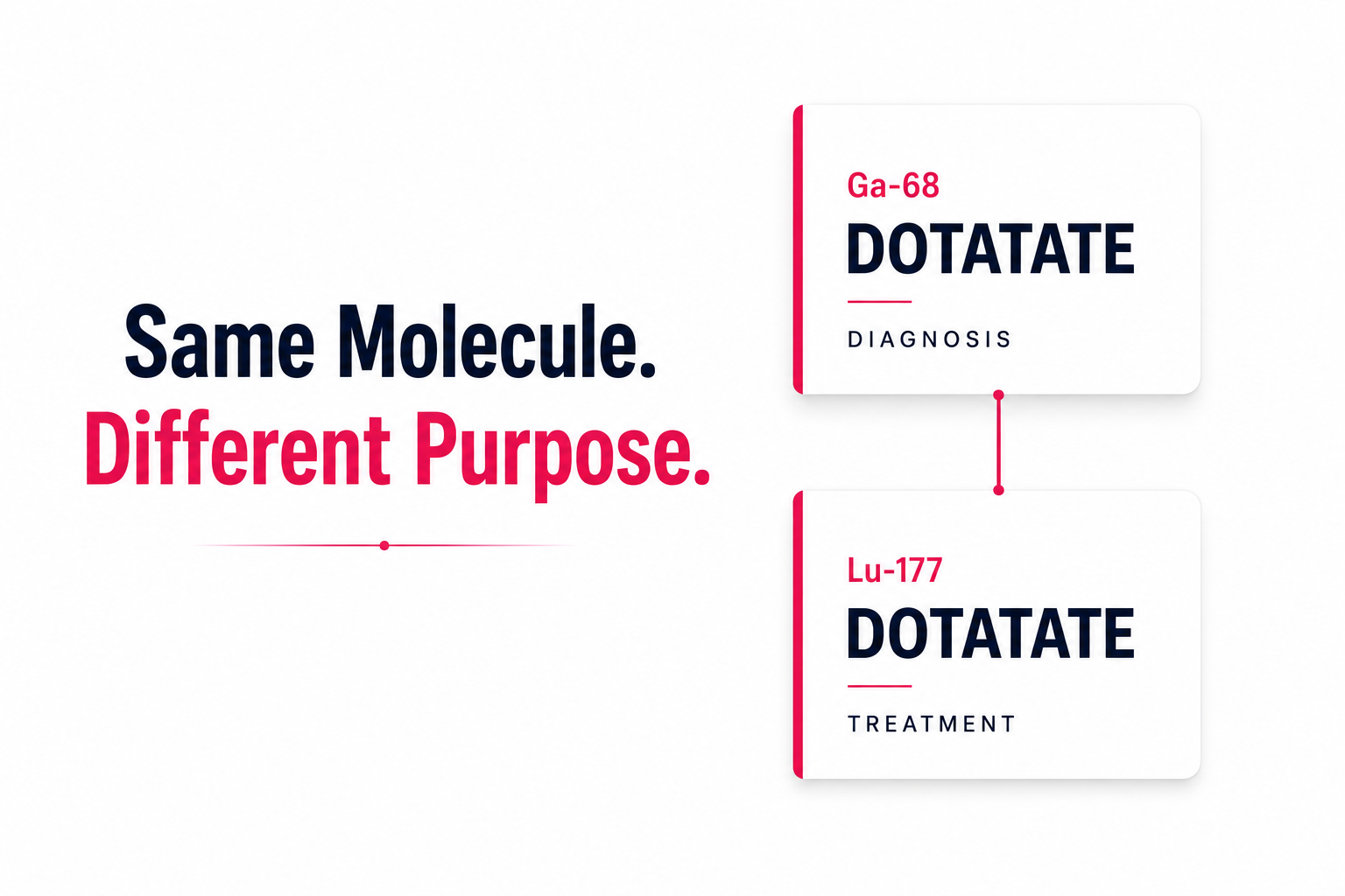
Neuroendocrine tumours (carcinoid tumours, gastrinomas, pheochromocytomas) frequently metastasise to the liver. FDG PET CT should be used for most lymphomas — but for neuroendocrine tumours, somatostatin-targeting peptides such as DOTANOC and DOTATATE are used instead.
For neuroendocrine liver metastases — FDG PET CT is frequently negative. Well-differentiated NETs have low glucose metabolism. The correct investigation is DOTANOC or DOTATATE PET CT — which maps somatostatin receptor expression on NET cells.
If your liver cancer is actually a NET liver metastasis — confirm with your oncologist whether DOTANOC PET CT is required rather than FDG PET CT. The page FDG vs PSMA vs DOTANOC → covers this distinction in detail.
The Triple Phase PET CT Connection
For certain liver cancer evaluations — particularly in patients being considered for liver-directed therapy — a Triple Phase PET CT may be ordered. This protocol combines FDG PET CT with a three-phase contrast CT of the liver (arterial, portal venous, and delayed phases) in a single examination.
This provides both:
- FDG metabolic activity mapping for whole body staging
- High-resolution triphasic liver CT for precise characterisation of hepatic lesions
The Triple Phase PET CT is particularly relevant for HCC patients where the arterial enhancement pattern of liver lesions is diagnostically important alongside metabolic activity.
Triple Phase PET CT cost in Delhi: ₹18,000–₹28,000 at AERB-licensed nuclear medicine centres. See full guide → Triple Phase PET CT Cost Delhi →
Portal Vein Thrombosis — A Unique HCC Application
PET CT can differentiate between benign and malignant portal vein thrombosis in HCC.
Portal vein thrombosis is common in patients with HCC and cirrhosis. Benign portal vein thrombosis from cirrhosis does not affect surgical candidacy in the same way as malignant portal vein invasion by tumour. FDG PET CT — by showing FDG uptake or absence in the thrombus — can help distinguish bland thrombus from tumour thrombus, with significant implications for treatment planning and transplant eligibility.
FDG PET CT Cost for Liver Cancer in Delhi 2026
|
Indication |
Tracer |
AERB-Licensed Centre (₹) |
Hospital (₹) |
CGHS Rate (₹) |
|
HCC extrahepatic staging / recurrence |
FDG |
9,000 – 14,000 |
25,000 – 38,000 |
21,000 |
|
Liver metastases from colorectal/breast/lung |
FDG |
9,000 – 14,000 |
25,000 – 38,000 |
21,000 |
|
NET liver metastases |
DOTANOC / DOTATATE |
16,000 – 22,000 |
28,000 – 42,000 |
17,000 |
|
Triple Phase PET CT |
FDG + triphasic CT |
18,000 – 28,000 |
35,000 – 50,000 |
Confirm |
All prices from EVE Healthcare partner centres — AERB-licensed nuclear medicine centres in Delhi NCR. Prices verified June 2026.
Pre-Procedure Considerations for Liver Cancer PET CT
For HCC patients on sorafenib or lenvatinib (targeted therapy): Targeted therapy affects tumour metabolism — FDG uptake patterns in treated tumours may differ from untreated lesions. Your oncologist will advise on optimal timing relative to your treatment cycle.
For HCC patients with cirrhosis: The background liver in cirrhosis has altered glucose metabolism that affects FDG distribution. The nuclear medicine physician interpreting your report is aware of this — but informing them of your cirrhosis and underlying hepatitis status at booking ensures the report is contextualised appropriately.
For NET patients: If you are on octreotide LAR or lanreotide (long-acting somatostatin analogues) and have been referred for DOTANOC PET CT — these must be paused 4–6 weeks before the scan. Confirm with your oncologist. Do not pause independently.
Standard FDG preparation: Low-carbohydrate diet 24 hours before. 6-hour fast. Water freely. Full guide → How to Prepare for PET CT
Delhi NCR Availability for Liver Cancer PET CT
|
Area |
FDG PET CT |
DOTANOC Available |
Triple Phase |
CGHS |
|
South Delhi (Green Park, Safdarjung) |
Yes |
Yes |
Select |
Yes |
|
Central Delhi (Karol Bagh) |
Yes |
Yes |
Select |
Yes |
|
North Delhi (Rohini) |
Yes |
Select |
Select |
Yes |
|
Noida (Sector 17, 62) |
Yes |
Select |
Select |
Yes (select) |
|
Gurgaon (Golf Course, Sector 38) |
Yes |
Select |
Select |
Yes (select) |
Clinical Note
From the reviewing nuclear medicine physician: The liver cancer PET CT consultation I have most often in Delhi is a hepatologist referring an HCC patient "for PET CT staging." My first question is always: is this for intrahepatic staging or extrahepatic staging? For intrahepatic HCC characterisation — the hepatologist needs a contrast-enhanced MRI, not FDG PET CT. FDG PET CT misses approximately half of well-differentiated HCCs — which is the predominant HCC type in Indian patients with hepatitis B. However, for the same patient being evaluated for liver transplantation or major resection — FDG PET CT is exactly the right investigation for ruling out occult bone metastases, extrahepatic lymph node disease, or lung deposits that would make the patient inoperable. These are two different clinical questions that require two different investigations from the same patient. The second most common liver cancer PET CT question I receive is from colorectal cancer patients referred for liver metastasectomy evaluation — these patients have well-differentiated colorectal liver metastases that are FDG-avid, and the PET CT accurately maps whether the liver is the only site of disease and whether the resection is technically feasible. For these patients FDG PET CT is the single most important pre-operative investigation. When you call EVE Healthcare to book a liver cancer PET CT — tell our team whether this is for primary HCC or for liver metastases from another cancer. The clinical question determines the investigation.
Book Your Liver Cancer PET CT in Delhi
When booking: tell our team whether this is for primary HCC or liver metastases from another cancer, and the tracer specified on your referral. We confirm the correct investigation and tracer availability before your appointment.
WhatsApp +91 9990032078 or use the search tool at eve-healthcare.com.
Also see: Whole Body PET CT Scan Cost Delhi → · FDG vs PSMA vs DOTANOC → · DOTANOC PET CT Scan Cost Delhi → · Triple Phase PET CT Cost Delhi → · How to Prepare for PET CT → · CGHS PET CT Rate Delhi