If you have been diagnosed with lymphoma and your oncologist has referred you for a PET CT scan — this page explains exactly what the scan will show, what the Deauville score on your report means, the three different stages of the lymphoma journey where PET CT is used, and what it costs in Delhi.
FDG PET CT for lymphoma costs ₹9,000–₹14,000 at AERB-licensed nuclear medicine centres in Delhi — the same scan at equivalent quality centres compared to ₹25,000–₹38,000 at private hospital radiology departments.
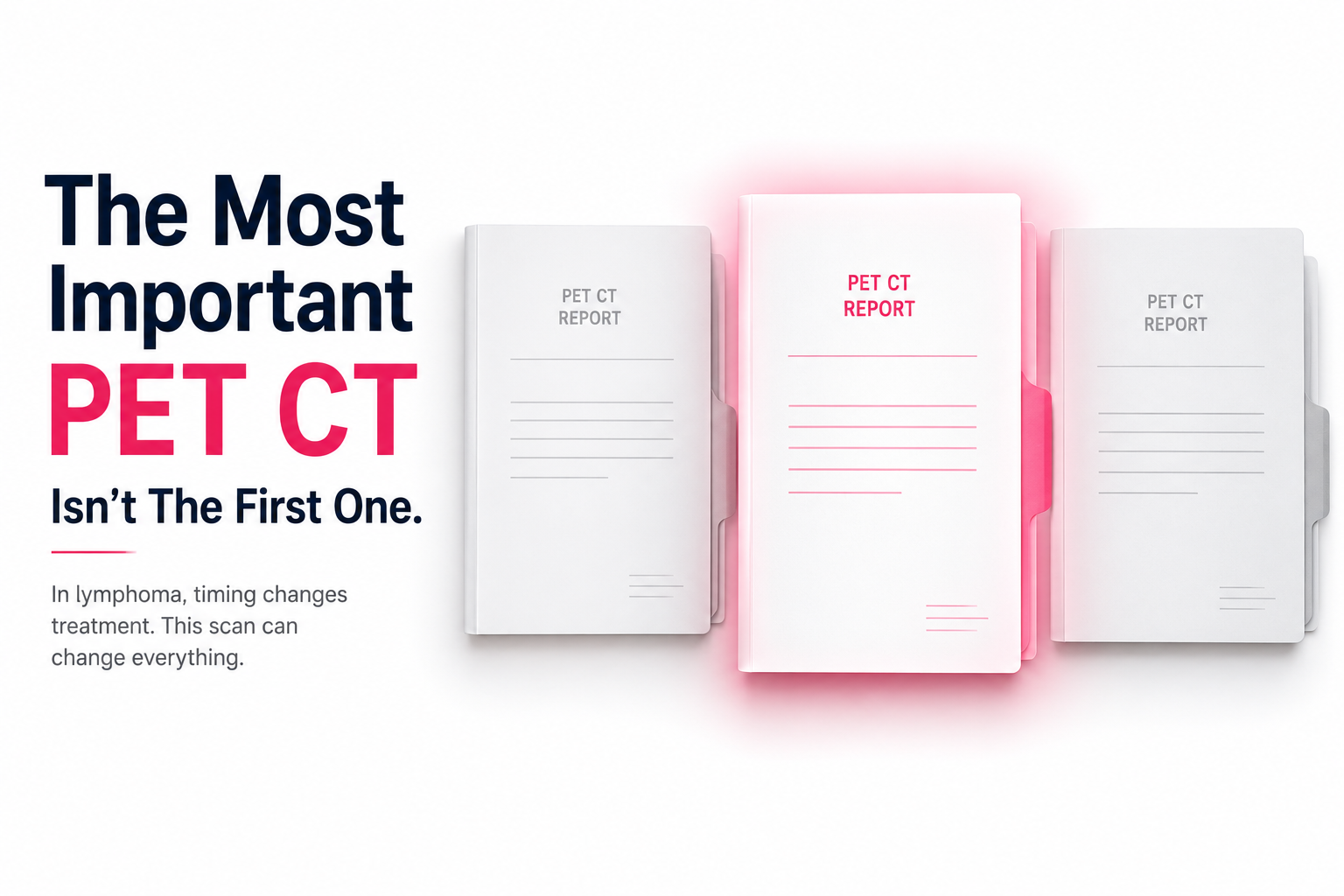
Why Lymphoma Uses PET CT More Than Any Other Cancer
Lymphoma has a unique relationship with FDG PET CT that goes beyond staging.
International guidelines recommend that PET CT be used for routine staging of FDG-avid lymphomas and for response assessment using the Deauville 5-point scale. American Migraine Foundation
FDG PET CT should be used for most lymphomas — including Hodgkin lymphoma, aggressive and high-grade Non-Hodgkin lymphomas such as diffuse large B-cell lymphoma, and many indolent lymphomas such as follicular lymphoma.
In most cancers, PET CT is used once or twice — for initial staging and possibly for restaging after treatment. In lymphoma, PET CT is used at every decision point in the treatment journey:
|
Stage of Journey |
When PET CT Is Used |
What It Determines |
|
Before treatment |
Initial staging — Ann Arbor/Lugano staging |
Stage I–IV, treatment regimen selection |
|
After 2 cycles of chemotherapy |
Interim PET CT (iPET) |
Whether treatment is working — may change regimen |
|
After completing treatment |
End-of-treatment PET CT |
Complete remission vs residual disease |
|
During surveillance |
Recurrence monitoring |
Whether remission is maintained |
This is why lymphoma patients become the most experienced PET CT patients in nuclear medicine — many will have 3–5 PET CT scans over their treatment course.
Which Lymphomas Use FDG PET CT?
Hodgkin lymphoma, all aggressive and many indolent Non-Hodgkin lymphomas should undergo FDG PET CT for staging and response assessment. In small lymphocytic lymphoma and chronic lymphocytic leukaemia, CT is used instead. Journal of Urgent Care Medicine
FDG PET CT is the standard for:
- Hodgkin Lymphoma (HL) — classical HL and nodular lymphocyte-predominant HL
- Diffuse Large B-Cell Lymphoma (DLBCL) — most common aggressive NHL
- Follicular Lymphoma (FL) — most common indolent NHL
- Mantle Cell Lymphoma
- T-cell lymphomas
FDG PET CT is NOT used for:
- Small Lymphocytic Lymphoma / Chronic Lymphocytic Leukaemia (SLL/CLL) — CT is used unless transformation to high-grade lymphoma is suspected
- Non-FDG-avid indolent NHLs — CT measures size changes
If you are unsure whether your lymphoma subtype uses PET CT — your haematologist or oncologist will specify on the referral.
What FDG PET CT Shows in Lymphoma — Stage by Stage

Initial Staging PET CT — The Baseline Scan
The baseline PET CT maps all FDG-avid disease sites in the body — lymph nodes above and below the diaphragm, the spleen, bone marrow (in Hodgkin's lymphoma specifically), and extranodal sites.
The Ann Arbor / Lugano Staging System:
|
Stage |
Disease Location |
Treatment Implication |
|
Stage I |
Single lymph node region or single extranodal site |
Early-stage — shorter chemotherapy ± radiotherapy |
|
Stage II |
Two or more regions on the same side of the diaphragm |
Early or intermediate stage |
|
Stage III |
Regions on both sides of the diaphragm |
Advanced stage |
|
Stage IV |
Widespread extranodal involvement (bone marrow, liver, lungs) |
Advanced stage — full chemotherapy course |
The PET CT advantage over CT alone: PET CT detects disease at extranodal sites — bone marrow involvement, splenic disease, and extranodal deposits — that CT misses because they are metabolically active without being enlarged. In Hodgkin's lymphoma specifically, FDG PET CT impacts staging with fewer cases with Stage I disease and more with skeletal involvement than CT staging alone. American Migraine Foundation
Bone marrow biopsy and PET CT: While FDG PET CT is sufficient to rule out bone marrow involvement in Hodgkin lymphoma, biopsy may be needed in other lymphomas. nih
For Hodgkin's lymphoma — if the PET CT shows no bone marrow FDG uptake, bone marrow biopsy can be avoided. For Non-Hodgkin's lymphomas, biopsy may still be required even with a negative PET CT.
Interim PET CT — The Most Consequential Scan
The interim PET CT — performed after 2 cycles of ABVD (for Hodgkin's) or R-CHOP (for DLBCL) — is the most clinically consequential scan in the entire lymphoma journey. Its result directly determines whether the current treatment continues, escalates, or de-escalates.
Interim FDG PET CT after two cycles of ABVD showed a negative predictive value of 93.6% for predicting treatment outcomes in Hodgkin's lymphoma — with Deauville score stratification into DS 1-3 versus DS 4-5 guiding treatment adaptation. Jacr
What this means: A negative interim PET CT (Deauville 1–3) has a 93.6% negative predictive value for unfavourable outcomes — meaning the vast majority of patients with a negative interim PET CT will achieve durable remission with continued standard treatment.
The treatment adaptation principle:
- Deauville 1–3 interim PET CT → treatment de-escalation may be considered (e.g. dropping bleomycin from ABVD in Hodgkin's, reducing cycles) — to reduce toxicity
- Deauville 4–5 interim PET CT → treatment escalation is considered (switching to more intensive regimen such as BEACOPPesc)
This is what makes the interim PET CT unique — it is not just diagnostic, it is treatment-adaptive. The same scan can lead two patients on the same initial regimen to completely different treatment pathways.
End-of-Treatment PET CT — The Remission Scan
After completing the full chemotherapy course, the end-of-treatment PET CT determines whether the patient has achieved complete metabolic remission.
Complete Metabolic Response (CMR): No FDG uptake above background at previously involved sites. Deauville 1–2. The treatment has worked. The patient is in complete metabolic remission.
Partial Metabolic Response (PMR): Reduced but persistent FDG uptake. Deauville 3 (often acceptable in certain situations) or 4–5 (concerning — further investigation needed).
The residual mass problem: Many lymphoma patients have residual masses on CT after completing treatment — enlarged nodes that have not completely resolved in size. On CT alone, a persistent mass after treatment raises concern for residual disease. On PET CT, if the mass is FDG-negative (Deauville 1–2), it represents fibrotic or necrotic tissue — not active disease. PET CT resolves the residual mass ambiguity that CT cannot.
The Deauville Score — What It Means and How to Read It
The Deauville score is the reporting standard for all lymphoma PET CT scans. Every response assessment PET CT report will include a Deauville score from 1 to 5.
For treatment response assessment, the 5-point Deauville scale that compares FDG uptakes of lymphoma manifestations and reference tissues on PET should be used. nih
|
Deauville Score |
What It Means |
Clinical Interpretation |
|
1 |
No FDG uptake above background |
Complete metabolic response — excellent |
|
2 |
FDG uptake ≤ mediastinal blood pool |
Complete metabolic response — excellent |
|
3 |
FDG uptake > mediastinal blood pool but ≤ liver |
Adequate response in most protocols — discuss with oncologist |
|
4 |
FDG uptake moderately > liver |
Inadequate response — treatment change likely discussed |
|
5 |
FDG uptake markedly > liver or new sites of disease |
No response or progression — treatment change indicated |
|
6 |
New areas of FDG uptake unlikely to be related to lymphoma |
Requires separate assessment |
What the reference points mean:
- Mediastinal blood pool: The blood in the large vessels of the chest — has a normal SUV of approximately 1.5–2.0
- Liver: Normal liver tissue has SUV of approximately 2.0–2.5
A Deauville 3 means the residual uptake in a treated site is between these two reference points — above the mediastinal blood pool but not above the liver. Whether this is acceptable depends on the specific lymphoma protocol and the clinical context.
The most important message about Deauville scores: Do not interpret your Deauville score without your oncologist. Deauville 3 in an interim scan after 2 cycles of ABVD has a very different management implication from Deauville 3 in an end-of-treatment scan. The same number, different clinical meaning.
The Baseline PET CT — The Most Important Instruction EVE Can Give
When you come for your interim or end-of-treatment PET CT — always bring your baseline PET CT report and images.
This is the booking instruction that prevents the most common lymphoma PET CT quality failure in Delhi.
The nuclear medicine physician reporting your interim or end-of-treatment PET CT needs your baseline images for side-by-side comparison. Deauville scoring requires comparing current FDG uptake at previously involved sites to what was present at baseline. Without the baseline images:
- The physician cannot confidently determine whether a residual FDG-avid site represents persistent disease or physiological uptake
- Deauville scoring is less precise — the physician must rely on memory or estimation rather than direct comparison
- The quality and clinical utility of the report is compromised
What to bring:
- CD or USB drive with baseline PET CT images (DICOM format — ask the original nuclear medicine centre for a copy)
- Printed baseline PET CT report
- Any previous interim PET CT reports if this is a later follow-up scan
When booking through EVE Healthcare for an interim or end-of-treatment lymphoma PET CT — tell our team it is a follow-up scan. We will remind you to bring your baseline images and flag this to the nuclear medicine centre before your appointment.
FDG PET CT Cost for Lymphoma in Delhi 2026
|
Option |
Cost (₹) |
Waiting Time |
Notes |
|
AERB-licensed standalone nuclear medicine centre |
9,000 – 14,000 |
Same-day to 3 days |
Equivalent machine quality |
|
Private hospital (Rajiv Gandhi Cancer, Max, Fortis) |
25,000 – 38,000 |
2–7 days |
Premium pricing |
|
Government centre (AIIMS, IRCH) |
3,500 – 6,000 |
3–8 weeks |
Long wait — not appropriate for interim or end-of-treatment scans with time-sensitive decisions |
|
CGHS-empanelled nuclear medicine centre |
21,000 (CGHS rate) |
Same-week |
CGHS beneficiaries only |
Lymphoma-specific cost reality: A typical Hodgkin's lymphoma patient on ABVD requires:
- 1 baseline PET CT (before treatment)
- 1 interim PET CT (after 2 cycles)
- 1 end-of-treatment PET CT (after 6 cycles)
- Possible surveillance PET CT if clinically indicated
Total: 3–4 FDG PET CT scans over treatment course.
At ₹10,000–₹14,000 per scan at a standalone nuclear medicine centre: ₹30,000–₹56,000 total. At ₹25,000–₹38,000 per scan at a private hospital: ₹75,000–₹1,52,000 total.
The cumulative cost difference over a lymphoma treatment course is ₹45,000–₹96,000 — for identical diagnostic quality.
Lymphoma PET CT Near Me — Delhi NCR
|
Area |
Price Range |
Same-Day FDG |
CGHS |
Metro Access |
|
South Delhi (Green Park, Safdarjung, Saket) |
₹9,000 – ₹14,000 |
Yes |
Yes (select) |
Yellow / Violet Line |
|
North Delhi (Rohini, Pitampura) |
₹9,000 – ₹13,000 |
Yes |
Yes |
Red Line |
|
Central Delhi (Karol Bagh) |
₹9,000 – ₹14,000 |
Yes |
Yes |
Blue / Yellow Line |
|
Noida (Sector 17, 62) |
₹9,000 – ₹13,000 |
Yes |
Yes (select) |
Blue Line |
|
Gurgaon (Golf Course Road, Sector 38) |
₹9,000 – ₹14,000 |
Yes |
Yes (select) |
Yellow Line |
Preparation for FDG PET CT — Lymphoma Patients on Active Treatment
Standard FDG preparation applies — but two lymphoma-specific points:
Steroid use: Many lymphoma patients receive steroids as part of their chemotherapy regimen (e.g. prednisone in CHOP). Inform the nuclear medicine physician of your steroid dose and timing before the scan. Steroids may affect FDG distribution in some protocols.
Timing relative to chemotherapy:
- Baseline PET CT: Before chemotherapy begins — no timing constraint
- Interim PET CT: Typically 2–3 weeks after the second chemotherapy cycle — to allow any treatment-related inflammatory FDG uptake to settle
- End-of-treatment PET CT: Typically 3–4 weeks after the last chemotherapy cycle
Your oncologist will specify the exact timing — follow their instruction rather than a general guideline.
Standard preparation:
- Low-carbohydrate diet 24 hours before the scan — no roti, rice, dal, fruit, or sugar
- Complete fast for 6 hours before injection
- Water freely throughout
- No strenuous exercise for 48 hours before
Full guide → How to Prepare for PET CT →
Clinical Note
From the reviewing nuclear medicine physician: Lymphoma is the lymphoma patient's disease in the truest sense — because these patients arrive for their second, third, and fourth PET CT already knowing what a Deauville score is, what an SUVmax means, and what a positive interim scan implies for their treatment. The conversations I have with lymphoma patients are different from any other oncology PET CT — they are informed, they ask precise questions, and they understand the stakes of each scan. What I tell every lymphoma patient coming for an interim PET CT in Delhi: bring your baseline images. Not the printed report — the actual DICOM images on a CD or USB. Without them, I am comparing your current scan to my memory of what a typical baseline looks like for your lymphoma subtype, rather than to your actual baseline. The precision of Deauville scoring depends on that comparison. A Deauville 3 that I call correctly on a side-by-side comparison might be called a Deauville 4 without the baseline — and in some protocols, that distinction changes whether your oncologist considers your treatment adequate or not. The CD is not a formality. It is a clinical tool.
Book Your Lymphoma PET CT in Delhi
Same-week availability at AERB-licensed nuclear medicine centres across Delhi, Noida, and Gurgaon. When booking for an interim or end-of-treatment scan — tell our team it is a follow-up and we will confirm the baseline image requirement before your appointment.
WhatsApp +91 9990032078 or use the search tool at eve-healthcare.com.
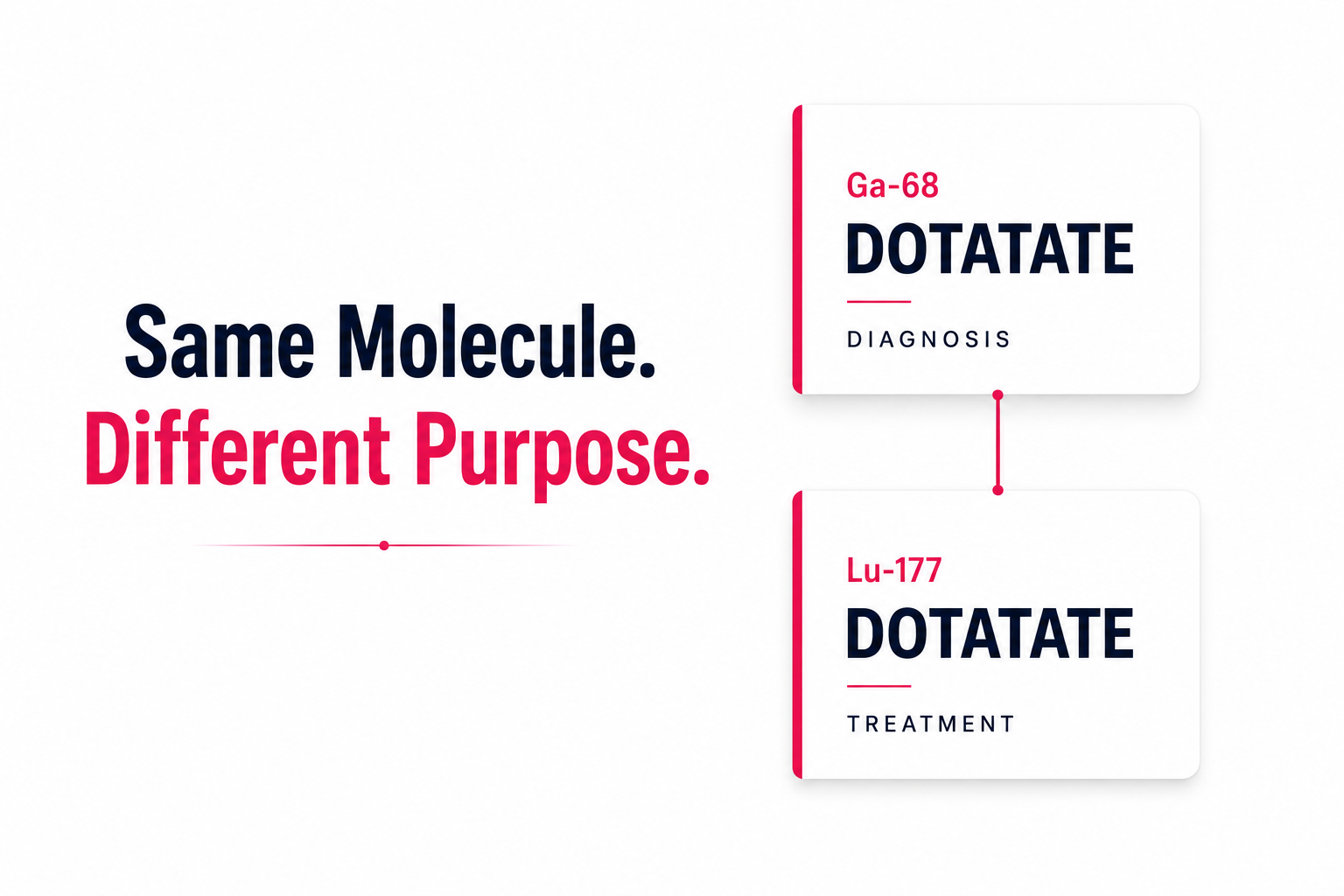
Also see: Whole Body PET CT Scan Cost Delhi → · FDG vs PSMA vs DOTANOC → · How to Prepare for PET CT → · What Is SUV Value? → · CGHS PET CT Rate Delhi → · PET CT Scan Lung Cancer Delhi →