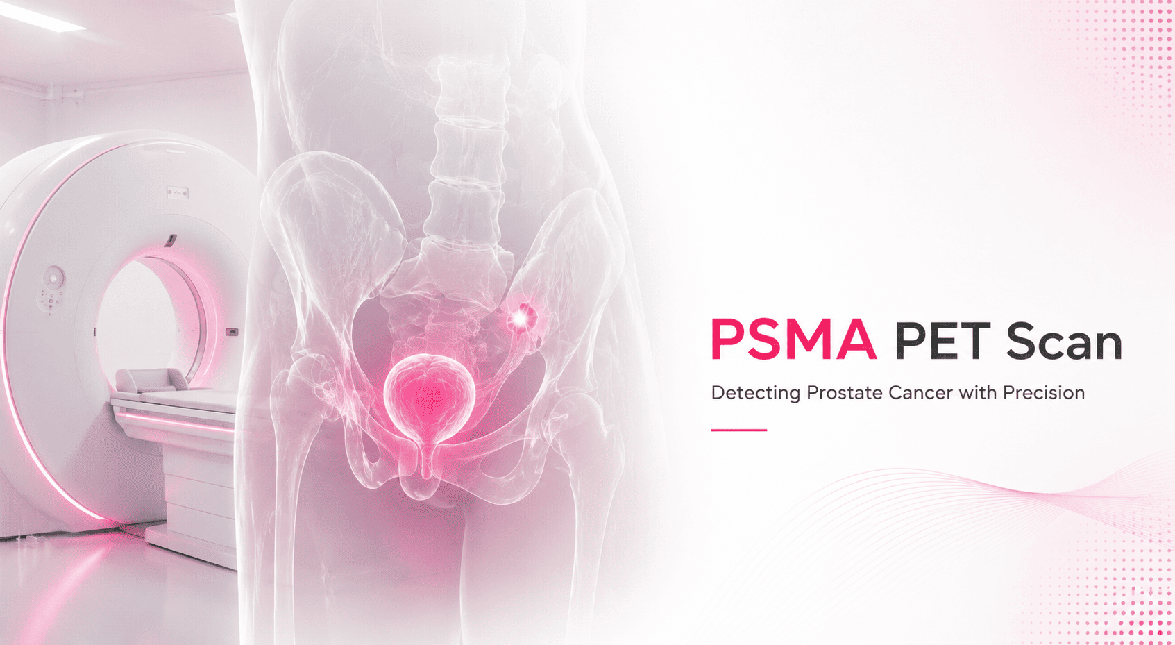
A PSMA PET scan has become the most accurate imaging available for prostate cancer, capable of finding disease at PSA levels where every older scan comes back blank. This guide explains how it works, when it is used, and how to make sense of the report, including the SUVmax number and the report scores that worry patients most.
If you are looking for pricing, see our PSMA PET CT scan cost in Delhi page. This page is about the scan itself.
How a PSMA PET Scan Works
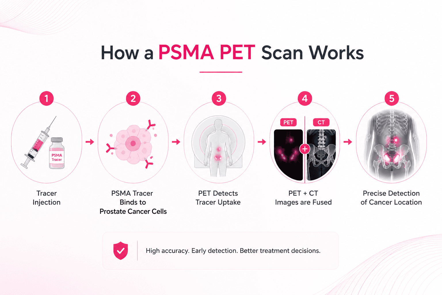
PSMA stands for Prostate-Specific Membrane Antigen, a protein expressed at high levels on the surface of most prostate cancer cells. A PSMA PET scan injects a small amount of a radioactive tracer engineered to bind to that protein. Wherever prostate cancer cells are, the tracer collects, and the PET scanner detects it as a bright signal.
The PET image is then fused with a CT scan, giving two layers at once: the PET layer showing tracer activity, usually in colour, and the CT layer showing anatomy in greyscale. This fusion is what lets a radiologist say not just that there is disease, but exactly which lymph node, which bone, or which part of the prostate bed it sits in.
The reason it has transformed prostate cancer imaging is sensitivity. PSMA tracers identify cancer at very low PSA levels, opening the door to early, targeted treatment of recurrence that would otherwise be invisible.
When Is a PSMA PET Scan Ordered?
Your urologist or oncologist orders a PSMA PET scan to:
- Stage moderate to high-risk prostate cancer at diagnosis, before deciding on treatment
- Find recurrence when PSA rises after surgery or radiotherapy, known as biochemical recurrence
- Detect metastases in lymph nodes and bone
- Assess treatment response
- Determine eligibility for PSMA-targeted radioligand therapy, such as Lutetium-177 PSMA
Its greatest value is at low PSA, where it detects disease that a conventional CT or bone scan simply cannot see.
How to Read a PSMA PET Report
This is what most people arrive here for, often holding a report full of numbers and unfamiliar terms.
SUVmax
The figure that draws the eye is SUVmax, the maximum standardized uptake value. It quantifies how intensely tracer accumulates in a given spot. Higher means stronger PSMA expression, which correlates with cancer.
To give a sense of scale from published data: an SUVmax above 2.5 flagged suspicious prostate lesions, high-grade tumours showed SUVmax above 12, and a cutoff of 5.4 separated low-grade from high-grade disease with 100 percent specificity in one study.
But a single number is not a diagnosis. Inflammation, normal organs, and benign structures can all take up tracer. SUVmax is read in context, against the pattern, the location, and your clinical picture.
PSMA-RADS and PROMISE
Because different readers could otherwise reach different conclusions, the field adopted standardized reporting systems. PROMISE assigns each suspected lesion a score from 0 to 3, comparing its uptake against reference organs, the blood pool, liver, and parotid gland, and applies a staging system called miTNM. PSMA-RADS grades how likely a finding is to be genuine prostate cancer.
If your report mentions these, it is a good sign: it means the scan was interpreted to a recognised standard, and the result is comparable across scans and centres.
What a Normal PSMA PET Scan Looks Like
A crucial point that alarms many patients: some bright areas are entirely normal.
Certain healthy organs always show high PSMA uptake, either because they express the protein physiologically or because the tracer is excreted through them. These include the salivary and lacrimal glands, liver, spleen, kidneys, small bowel, and bladder. Seeing intense signal there does not mean cancer has spread to them.
Normal or benignly enlarged prostate tissue shows only low to moderate uptake. Cancer typically appears as focal uptake standing clearly above the surrounding normal tissue. Distinguishing the two is precisely the skill of the nuclear medicine physician reading your scan, which is why who reads it matters as much as the scan itself.
Gallium-68 vs Fluorine-18 PSMA — Which Tracer?
You may see your scan described as Ga-68 PSMA or F-18 PSMA. Both target the same protein and do the same job. They differ in the radioactive label attached.
Gallium-68 PSMA-11 is generator-produced, widely established, and cleared largely through the urinary tract.
Fluorine-18 tracers, such as F-18 PSMA-1007 and F-18 DCFPyL, offer practical advantages: they are made in larger batches, so availability is better, they give higher image resolution, and their longer half-life allows transport between centres.
Diagnostic performance is broadly comparable. The differences are in biodistribution and logistics. F-18 PSMA-1007, for instance, shows more benign bone uptake that an inexperienced reader could mistake for metastases, while Gallium-68 clears more through the bladder. In practice, which tracer you receive often depends on what your centre has available that day.
PSMA PET vs Bone Scan and Other Imaging
For bone metastases, PSMA PET is generally more sensitive than a conventional bone scan. In one comparison of 32 patients, Gallium-68 PSMA PET/CT found more bone metastases than an F-18-NaF bone scan in 20 patients, including lesions the bone scan missed entirely in 7. PSMA PET also assesses lymph nodes and soft tissue in the same study, which a bone scan cannot.
For choosing between PSMA PET and other PET tracers, see our guide on FDG vs PSMA vs DOTANOC vs DOPA PET CT.
The Honest Limitations
PSMA is not perfect, and knowing its limits prevents unnecessary alarm.
It is not entirely specific. Uptake can occur in benign bone lesions, ganglia, inflammation, and some non-prostate tumours. This is exactly why PSMA-RADS exists and why an experienced reader is essential.
Occasionally, a patient with a rising PSA has a negative PSMA scan. In that situation, other tracers such as F-18 fluciclovine or choline may be considered, because a small proportion of prostate cancers express little PSMA.
A positive finding is a reason for careful interpretation and discussion with your oncologist, not for panic based on the SUV number alone.
Related Guides
- PSMA PET CT Scan Cost in Delhi
- FDG vs PSMA vs DOTANOC vs DOPA PET CT
- PET CT for Prostate Cancer in Delhi
- Is PET CT Scan Safe?
EVE Healthcare arranges PSMA PET CT at AERB-licensed nuclear medicine centres across Delhi NCR, subject to tracer availability, with reports read by nuclear medicine physicians. WhatsApp +91 9990032078 or call.