MRI Chest: A Detailed Overview
Introduction
An MRI of the chest (also known as a thoracic MRI) is a non-invasive diagnostic imaging technique that provides detailed images of the structures within the chest, including the lungs, heart, blood vessels, esophagus, lymph nodes, and chest wall. Unlike X-rays or CT scans, MRI uses a powerful magnetic field and radio waves to produce images without the use of ionizing radiation, making it a safer option for certain patients.
In this blog, we'll explore the indications for a chest MRI, the procedure involved, common conditions it helps diagnose, and what patients can expect during the process.
What is MRI Chest Used For?
A chest MRI is typically recommended when physicians need detailed imaging of the soft tissues in the chest. It is often used when other imaging modalities, such as X-rays, CT scans, or ultrasound, do not provide sufficient information. Some common reasons for a chest MRI include:
-
Cardiac Evaluation:
- To assess heart conditions, such as cardiomyopathies, heart valve disease, and congenital heart defects.
- To visualize the heart’s structure, function, and blood flow.
-
Tumor Detection:
- To evaluate for tumors or masses in the chest wall, lungs, or mediastinum (the area between the lungs).
- To determine if a tumor is benign or malignant.
-
Blood Vessel Abnormalities:
- To identify abnormalities in major blood vessels, such as aortic aneurysms, dissections, or pulmonary artery abnormalities.
- It can also be used to evaluate vascular malformations or thrombosis (blood clots).
-
Chest Wall Conditions:
- To investigate chest wall deformities, infections, or trauma.
- Useful in detecting the spread of cancer to the chest wall.
-
Lymph Nodes:
- To examine enlarged lymph nodes, often associated with cancers, infections, or autoimmune diseases.
-
Lung Evaluation:
- Although CT is preferred for evaluating the lungs, MRI may be used to assess diseases affecting the pleura (the lining of the lungs), chest wall masses, or diaphragm abnormalities.
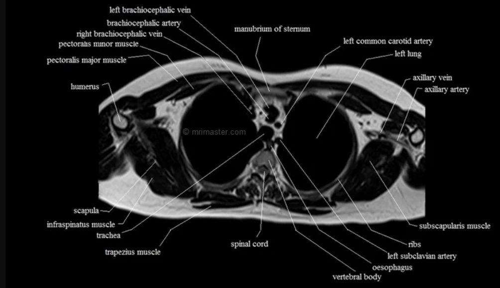
Anatomy of the Chest
The chest (thoracic cavity) houses vital structures, including:
- Heart: The central organ of the circulatory system that pumps blood throughout the body.
- Lungs: Organs responsible for the exchange of oxygen and carbon dioxide.
- Aorta and Major Blood Vessels: The large arteries and veins that carry blood to and from the heart.
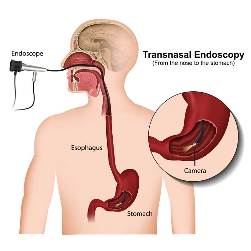
- Esophagus: The muscular tube that connects the throat to the stomach.
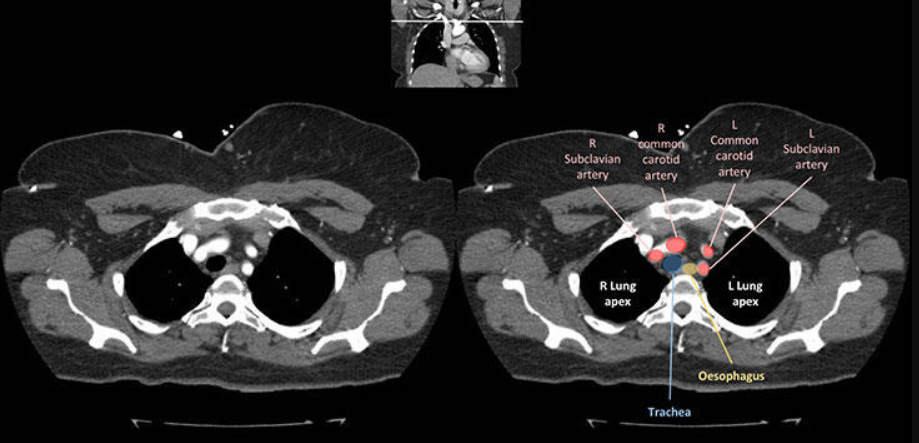
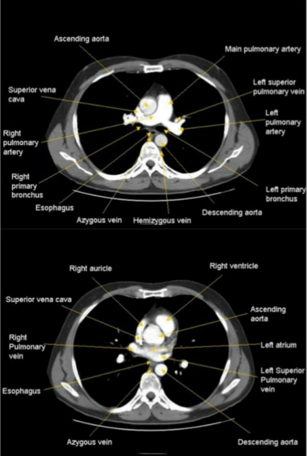
- Mediastinum: The area between the lungs that contains the heart, trachea, esophagus, and large blood vessels.
- Pleura: The thin membrane lining the lungs and chest cavity.
- Diaphragm: The muscle that separates the chest from the abdomen and plays a major role in breathing.
MRI Chest Procedure
1. Preparation for the Scan
- Medical History: Prior to the scan, you’ll be asked about any metal implants (like pacemakers, stents, or metal fragments), as MRI uses a magnetic field that can interfere with metal objects.
- Fasting: If a contrast agent is to be used, you may be instructed to avoid food and drink for a few hours before the exam.
- Clothing and Jewelry: You’ll need to remove metal objects, including jewelry, watches, and clothing with metal zippers or buttons. You’ll be asked to wear a hospital gown during the procedure.
2. During the Procedure
- Positioning: You will lie flat on a table that slides into the MRI machine. The machine resembles a large tube with an opening at both ends.
- Contrast Agent: In some cases, a contrast agent (usually gadolinium) will be injected into your vein to enhance the visibility of certain structures, such as blood vessels or tumors. The contrast is generally safe, though it may cause mild side effects like a metallic taste or warmth.
- Imaging Sequences: The scan involves multiple imaging sequences, each lasting several minutes. During each sequence, the machine will emit loud knocking or buzzing sounds. You’ll be provided with earplugs or headphones to block the noise.
- Breath-Holding: For clear images, you may be asked to hold your breath for short periods during the scan. This is particularly important for chest MRI since movement of the chest can blur the images.
3. After the Scan
- Completion: The procedure typically lasts between 30 minutes to an hour. Once it's complete, you can resume normal activities immediately unless you’ve been given a sedative.
- Results: A radiologist will analyze the MRI images and send a report to your doctor, who will discuss the findings with you.
Common MRI Chest Sequences
-
T1-Weighted Imaging: Offers detailed images of chest structures and is useful in evaluating tumors and abnormal tissues.
-
T2-Weighted Imaging: Highlights fluid-filled areas, making it ideal for detecting infections, inflammation, and edema.
-
STIR (Short Tau Inversion Recovery): Suppresses fat signals and is used to evaluate lymph nodes, muscles, and inflammation in the chest wall.
-
Contrast-Enhanced MRI: Utilizes gadolinium contrast to enhance the visibility of blood vessels, tumors, and areas of inflammation or scar tissue.
Conditions Diagnosed with Chest MRI
-
Cardiovascular Diseases:
- Myocarditis (inflammation of the heart muscle).
- Aortic aneurysms and dissections.
- Congenital heart defects.
- Pericardial effusion (fluid around the heart).
-
Tumors and Masses:
- Lung cancer (in some cases, though CT is preferred).
- Mediastinal tumors (tumors in the central chest area).
- Chest wall tumors (soft tissue or bone).
-
Chest Wall Conditions:
- Pectus excavatum (a chest wall deformity).
- Infections or trauma.
-
Vascular Abnormalities:
- Pulmonary embolism (blood clot in the lungs).
- Thrombosis (blood clot) in the chest’s major veins.
-
Lung and Pleura Diseases:
- Pleural effusion (fluid accumulation in the lung lining).
- Diaphragmatic paralysis.
Advantages of MRI Chest
-
No Radiation: MRI does not use ionizing radiation, making it a safer option for patients who need multiple imaging tests or are sensitive to radiation exposure.
-
Superior Soft Tissue Contrast: MRI is excellent for imaging soft tissues, such as the heart, blood vessels, and chest wall, which can be challenging to assess with other imaging techniques.
-
Multiplanar Imaging: MRI can produce images in multiple planes (axial, coronal, sagittal), providing more detailed information compared to X-rays or CT scans.
-
High-Resolution Imaging: MRI provides high-resolution images, making it ideal for detecting small lesions or abnormalities.
Risks and Limitations
-
Metal Implants: Patients with metal implants, such as pacemakers or cochlear implants, may not be eligible for MRI as the magnetic field can interfere with these devices.
-
Contrast Reaction: While rare, some patients may have an allergic reaction to the gadolinium contrast agent. It’s important to inform your doctor of any allergies or kidney problems before the scan.
-
Claustrophobia: The enclosed space of the MRI machine can cause anxiety in some patients. Open MRI machines or sedation can be offered to those who feel claustrophobic.
Conclusion
An MRI of the chest is a versatile and valuable imaging tool that helps diagnose a wide range of conditions affecting the heart, lungs, blood vessels, and chest wall. With its ability to produce detailed, high-resolution images without the use of radiation, MRI is an excellent choice for patients requiring comprehensive chest evaluations.
At EVE Healthcare, we offer cutting-edge MRI technology and experienced radiologists to ensure accurate diagnoses and optimal patient care. Whether you're being evaluated for heart disease, lung conditions, or chest wall abnormalities, our team is here to provide you with the best diagnostic imaging experience.
Contact Us
If you have questions about chest MRI or would like to schedule an appointment, please contact EVE Healthcare. We are committed to providing top-quality healthcare with a focus on patient safety and comfort.

.png)
.png)