MRI of the Sacrum and Coccyx (Tailbone): A Complete Guide
Introduction
The sacrum and coccyx (commonly referred to as the tailbone) are located at the base of the spine, playing a crucial role in supporting the upper body and connecting the spine to the pelvis. While these structures are relatively small compared to the rest of the spine, they can be involved in various conditions that cause significant discomfort or pain, such as trauma, fractures, tumors, or degenerative changes.
An MRI of the sacrum and coccyx is a highly sensitive and non-invasive imaging tool used to evaluate these structures, providing detailed images of both bone and soft tissues. This blog explores the purpose of sacrum and coccyx MRI, the procedure, common conditions it helps diagnose, and what to expect during the scan.
What is an MRI of the Sacrum and Coccyx?
A Magnetic Resonance Imaging (MRI) of the sacrum and coccyx utilizes powerful magnetic fields and radio waves to generate highly detailed images of these structures. This imaging modality is particularly useful for evaluating soft tissues, nerve roots, ligaments, and cartilage around the sacrum and coccyx, which may not be as clearly visible on X-rays or CT scans.
Indications for MRI of the Sacrum and Coccyx:
- Persistent pain in the lower back, buttocks, or tailbone area
- Coccygodynia (pain in the coccyx region)
- Trauma or injury to the sacrum or coccyx
- Fractures of the tailbone
- Tumors or abnormal growths near the sacrum or coccyx
- Infections or inflammatory conditions
- Congenital anomalies
- Degenerative diseases affecting the lower spine
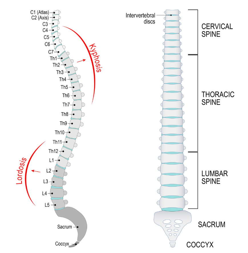
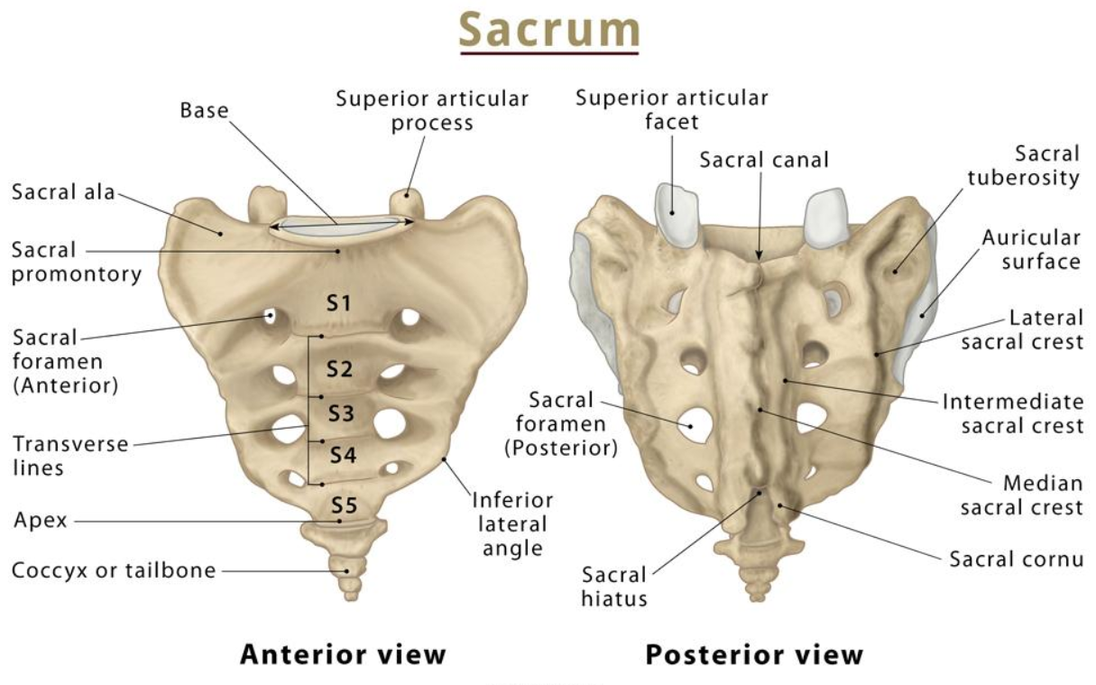
Anatomy of the Sacrum and Coccyx
-
Sacrum: A triangular-shaped bone located at the base of the lumbar vertebrae and connected to the pelvis. It is formed by the fusion of five vertebrae (S1-S5) and serves as the backbone of the pelvis.
-
Coccyx: Commonly known as the tailbone, the coccyx is made up of three to five small, fused vertebrae located below the sacrum. Though small, the coccyx provides attachment for ligaments, tendons, and muscles that play a role in supporting and stabilizing the pelvic region.
Why is an MRI of the Sacrum and Coccyx Performed?
MRI is often the preferred imaging modality when assessing the sacrum and coccyx for a variety of conditions. Here are some common reasons for performing this scan:
-
Trauma or Fractures: MRI is used to assess fractures in the sacrum or coccyx that may result from falls, accidents, or direct trauma. These fractures can sometimes be missed on X-rays, making MRI a better option for diagnosis.
-
Coccygodynia: Chronic tailbone pain, known as coccygodynia, is often difficult to diagnose using other imaging techniques. MRI provides detailed images of soft tissues around the coccyx, making it easier to identify possible causes such as inflammation, ligament damage, or misalignment.
-
Tumors and Masses: MRI is highly sensitive for detecting both benign and malignant tumors in the sacrum and coccyx, such as chordomas (rare malignant tumors that grow in the bones of the spine), metastases, or nerve sheath tumors.
-
Infections: MRI is excellent at identifying infections in the bone (osteomyelitis) or surrounding soft tissues. It can provide important information about the extent and severity of the infection.
-
Degenerative Conditions: Age-related changes such as degenerative disc disease or sacroiliitis (inflammation of the sacroiliac joints) can be evaluated with an MRI, which shows detailed images of both bone and soft tissues.
-
Congenital Abnormalities: Some patients may be born with structural abnormalities of the sacrum or coccyx. MRI is used to detect these abnormalities and plan appropriate treatment if needed.
-
Pelvic Floor Disorders: In cases where pelvic floor dysfunction is suspected, MRI can help visualize the relationship between the sacrum, coccyx, and surrounding muscles, ligaments, and nerves.
How is an MRI of the Sacrum and Coccyx Performed?
1. Preparation for the Scan
-
Medical History: Your doctor will review your medical history to ensure MRI is safe, particularly if you have any metal implants or devices, such as a pacemaker.
-
Remove Metal Items: You’ll be asked to remove any metal objects (jewelry, watches, belts) before the scan, as they can interfere with the magnetic field.
-
Fasting: If a contrast agent is required, you may be asked to fast for several hours before the scan.
2. During the MRI Procedure
-
Positioning: You will lie face-up on a sliding table that will move into the MRI machine. You must remain still during the scan to ensure clear images.
-
Contrast Injection: In some cases, a contrast agent (like gadolinium) may be injected into your bloodstream to enhance the images, particularly if tumors or infections are suspected.
-
Scan Duration: The procedure typically takes 30 to 45 minutes, depending on whether contrast is used and the number of images required.
-
Noise: MRI machines can be loud, producing knocking or tapping sounds. You will be provided with earplugs or headphones to make the experience more comfortable.
3. Post-MRI
-
After the Scan: You can resume your normal activities immediately after the MRI unless you’ve been sedated for anxiety. If a contrast agent was used, you’ll be advised to drink plenty of fluids to flush it from your body.
-
Results: A radiologist will interpret the MRI images and provide a report to your doctor, who will discuss the findings and recommend further action or treatment if necessary.
Key MRI Sequences for Sacrum and Coccyx Imaging
-
T1-Weighted Imaging:
- Offers high-resolution images of the bony structures of the sacrum and coccyx, making it useful for identifying fractures or tumors.
-
T2-Weighted Imaging:
- Highlights fluid and soft tissues, helping to detect inflammation, nerve compression, or soft tissue injuries.
-
STIR (Short Tau Inversion Recovery):
- Suppresses fat signals and enhances the visualization of soft tissue abnormalities, particularly for detecting inflammation, infections, or tumors.
-
Fat-Suppressed T2 Imaging:
- Used to better visualize abnormalities in the soft tissues, including ligament damage or inflammation around the sacrum and coccyx.
-
Contrast-Enhanced Imaging:
- If a contrast agent is used, it can help highlight abnormal blood flow in tumors, infections, or areas of inflammation.
Common Conditions Diagnosed with MRI of the Sacrum and Coccyx
-
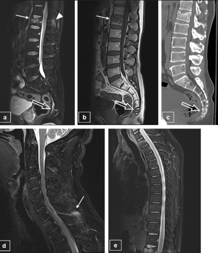
Coccygeal Fractures: MRI can detect fractures of the coccyx, which may result from falls or direct trauma. These fractures are often missed on X-rays.
-
Coccygodynia: Chronic pain in the tailbone region can be caused by a variety of conditions, including inflammation, trauma, or ligament damage. MRI is the best tool for identifying these causes.
-
Chordomas: These are rare tumors that can occur in the sacrum or coccyx. MRI is highly sensitive in detecting chordomas and evaluating their size and extent.
-
Sacroiliitis: Inflammation of the sacroiliac joints, often associated with conditions like ankylosing spondylitis, can cause severe lower back pain. MRI can detect inflammation in these joints.
-
Osteomyelitis: Infections in the bones of the sacrum or coccyx are best visualized using MRI, which can detect both bone and soft tissue involvement.
-
Degenerative Changes: Age-related wear and tear in the lower spine, including degenerative disc disease or arthritis, can be detected with MRI.
-
Pelvic Floor Dysfunction: MRI is sometimes used to evaluate the relationship between the coccyx and the muscles and ligaments of the pelvic floor, particularly in patients with pelvic pain or dysfunction.
Risks and Considerations
-
Claustrophobia: Some patients may feel anxious or claustrophobic in the enclosed space of an MRI machine. Open MRI machines or sedation options can be used to alleviate anxiety.
-
Metal Implants: Patients with metal implants, pacemakers, or certain medical devices may not be able to undergo an MRI. Always inform your doctor if you have any metal in your body.
-
Contrast Reactions: In rare cases, patients may experience an allergic reaction to the contrast agent. If you have kidney problems, inform your doctor, as contrast can sometimes affect kidney function.
Conclusion
An MRI of the sacrum and coccyx is a powerful diagnostic tool that provides detailed images of the tailbone, surrounding nerves, and soft tissues. Whether you’re experiencing chronic tailbone pain, trauma, or have been diagnosed with a condition affecting the sacrum or coccyx, an MRI can help identify the root cause and guide effective treatment.
At EVE Healthcare, we use advanced MRI technology to ensure accurate and precise diagnoses. Our experienced team is committed to delivering the highest quality care for your spine and tailbone concerns. If you’ve been recommended for a sacrum or coccyx MRI, rest assured that you’re in expert hands at EVE Healthcare.


.png)
.png)